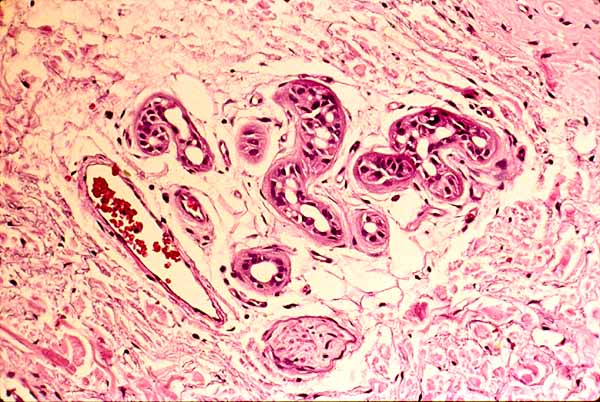

Sweat Gland in Skin

The portion of a sweat gland found deep in skin is surrounded by fibrous connective tissue of the dermis.
Sweat glands typically appear as clusters of several round or oval profiles, as shown here. Each profile represents a section across the twisted tubule which comprises the gland.
Point-and-click on the image above to identify sweat gland profiles and other structures (nerve, blood vessels, adipocytes).
Most of the area of this this image is occupied by collagen (pink), ground substance (pale background), and scattered fibroblasts and other connective tissue cells (dark).
To view this region in a larger context (low magnification) click here or on the thumbnail at right.
Comments and questions: dgking@siu.edu
SIUC / School
of Medicine / Anatomy / David
King
https://histology.siu.edu/intro/IN003b.htm
Last updated: 12 June 2022 / dgk