
Skin, dermis and hypodermis
Notes
Click here to view an interactive image with additional details labelled.
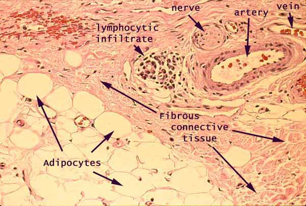
Subcutaneous adipose tissue occupies the lower left portion of the image. Fibrous connective tissue of the dermis occupies the remaining regions and includes a conspicuous artery, a small nerve, and a cluster of lymphocytes. Several smaller blood vessels may also be seen. Numerous capillaries in this area cannot be reliably recognized in this specimen.
The pink color in this image represents eosinophilic material, in this case mostly one of the following:
- Most of the pink material represents extracellular collagen fibers.
- The somewhat deeper pink encircling the artery at upper right represents the cytoplasm of smooth muscle cells.
The pale background color represents unstained material, in this case one of the following:.
- In the lower right, the large open spaces represent the lipid droplets within adipocytes. (Intact adipocytes appear round, with continuouss margins. Adipocytes which have been damaged during specimen preparation appear as irregular shapes with broken margins.)
- Within the vein and the artery, the pale space represents blood plasma (or, more accurately, the lumen where blood would flow in living tissue).
- Elsewhere, the thin interconnected pale spaces between collagen fibers represent connective tissue ground substance.
The very dark purple (nearly black) spots are cell nuclei. At this resolution, they cannot be individually identified except by context.
- Nuclei adjacent to the lumen of a blood vessel belong to vascular endothelial cells.
- Nuclei in the muscular wall of an artery may belong to smooth muscle cells.
- Irregular nuclei scattered at random among collagen fibers belong mostly to fibroblasts. Some may also belong to macrophages, mast cells, or capillary endothelial cells.
- Thin nuclei adjacent to large lipid droplets may belong to adipocytes or to fibroblasts or to capillary endothelium located between the adipocytes.
Bright red spots at upper left are red blood cells.
Blood is routinely spilled when tissue samples are taken. Usually, liquid blood is rinsed away during specimen preparation, so that even blood vessels themselves commonly appear empty. In this case, some spilled blood appears at upper left, and a few other blood cells appear in several of the blood vessels.
Comments and questions: dgking@siu.edu
SIUC / School
of Medicine / Anatomy / David
King
https://histology.siu.edu/intro/IN009b1.htm
Last updated: 22 May 2022 / dgk