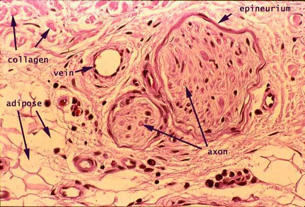

Skin, peripheral nerves in dermis

Notes
Nerves traveling through connective tissue deep in skin are larger and more readily recognized than those closer to epidermis.
A pair of small peripheral nerves are conspicuous features in this image, outlined by their epineurium. A few small blood vessels are also visible. A small amount of subcutaneous adipose tissue and fibrous connective tissue of the dermis occupy the remaining area.
Only the largest, myelinated axons are readily visible in routine light micrographs. Although few axons are identifiable in the nerves illustrated above, each nerve surely contains scores of smaller axons.
- In skin, large myelinated axons generally serve the sense of touch.
- Unmyelinated axons are associated with pain sensation and with autonomic motor function (controlling vascular smooth muscle and sweat glands).
The very dark purple (nearly black) spots are cell nuclei. At this resolution, most cannot be individually identified except by context.
- Nuclei within the nerves may be either Schwann cells or fibroblasts of the endoneurium.
- Nuclei adjacent to the lumen of a blood vessel belong to vascular endothelial cells.
- Irregular nuclei scattered at random among collagen fibers belong mostly to fibroblasts. Some may also belong to macrophages, mast cells, or capillary endothelial cells.
- A cluster of lymphocytes appear below the word "axon."
Comments and questions: dgking@siu.edu
SIUC / School
of Medicine / Anatomy / David
King
https://histology.siu.edu/intro/IN013b.htm
Last updated: 13 June 2022 / dgk