
Looking at blood smears
Interpreting blood smears is a specialized art. This page will not instill that art (much blood work is now done by machine, anyway) but rather provides only an initial introduction.
You may skip over the instruction on this page and go directly to sample blood smears.
The phrase "blood smear" is descriptive of how the preparation is made. A drop of blood is placed on the slide, smeared across it, fixed, dried, stained (commonly with Wright's stain; the name commemorates James Homer Wright, b. 1869), and covered. One can make smears not only of peripheral blood (circulating blood, taken from a vein) but also of bone marrow (where blood is formed) or of inflammatory infiltrate (pus).
When examining a blood smear, such as a slide from your reference collection, you should focus attention on suitable areas. The best region is typically toward the center of the smear, away from the edges.
(The following images were taken from a specimen from a sickle-cell patient. Due to inexpert image processing, color may vary from image to image.)
Avoid areas that resemble Figure 1, in which the blood cells are clumped or unevenly dispersed.
Also avoid areas which resemble Figure 2, where the blood cells are jumbled together and touching one another.
Concentrate your examination in areas where the cells are individually separated from one another and fairly uniformly dispersed, as in Figure 3.
Note that this latter image includes very many RBCs (including several with odd shapes, the "sickle cells") and two WBCs. The larger WBC (the one with the odd-shaped nucleus) is a neutrophil. The smaller WBC (the one with a round nucleus that mostly fills the cell) is a lymphocyte.
When viewing a blood smear, notice first the numerous red blood cells, pale cells without nuclei. Do not concentrate on individual cells, but scan over a large population of cells. Pay attention to the following features.
Typical cell size. Quantitative estimation requires special tools.
Variation in cell size. Are all cells nearly the same? Are many cells noticably bigger, or smaller, than the typical cell? Are there cells in both bigger and smaller categories?
Variation in cell shape. Do many cells appear in any shape that is dramatically different from round, such as pointy?
Variation in cell texture. Do all cells stain similarly pink, or do some contain basophilic patches or granules? (Recently-formed RBCs may retain bits of immature cytoplasm. The proportion of such cells is indicative of the rate at which RBCs are being produced.)
Next scan for white blood cells. These are larger than RBCs, with prominent blue (stained) nuclei. List by type each example as it is seen. By the time you have counted a hundred or so, you should have a fair estimate of the proportion of the commonest types.
In normal blood, a majority will be neutrophils and most of the remainder will be lymphocytes. These two types are easy to learn, because they are so common and because they look so different from one another.
Monocytes, eosinophils and basophils are a bit harder, partly because several hundred leukocytes (in a normal smear) must be examined to find and estimate these cell populations and partly because the range of appearance for these cells (especially monocytes) overlaps that for neutrophils and basophils.
Platelets are tiny (much smaller than RBCs) flecks of granular cytoplasm, often appearing in clumps.
A thorough blood smear examination includes not only the characteristics and proportions of mature cells but also the proportion of each type which appears immature.
Each blood cell type has several named maturational stages. Although most blood cell maturation occurs in bone marrow where the cells form, some immature forms also appear in peripheral blood.
A red cell which contains basophilic strands or granules (remnants of cytoplasm not yet completely replaced by hemoglobin) is called a reticulocyte.
Neutrophils are commonly identified as "bands" (immature, with a band-shaped nucleus) and "segs" (mature, with a nucleus segmented into distinct lobes).
Below is an example from a sickle-cell patient. Note variation in RBC size and shape. A key to the WBCs is below the image.
This micrograph does NOT represent an ideal sample from the patient's blood. Rather, it has been chosen to show how white blood cells can appear when artefactually damaged during preparation. Such unclassifiable blobs are not uncommon in ordinary blood smears.
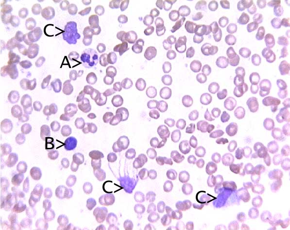
A. Neutrophil, segmented.
B. Lymphocyte.
C. Remains of damaged cells, to be ignored.Click here to view some sample blood smears.
For more information about blood cell types, including more images, see Blood cells. For more general information, return to Introduction to blood.
Comments and questions: dgking@siu.edu
SIUC / School
of Medicine / Anatomy / David
King
https://histology.siu.edu/intro/bldsmear.htm
Last updated: 13 November 2022 / dgk