Normal skin 
Insect bite (pathology report)

Melanoma (pathology report)

Healing wound (pathology report)


Examples of skin pathology
- Click here for basic orientation to skin.
- Click here for basic orientation to inflammation.
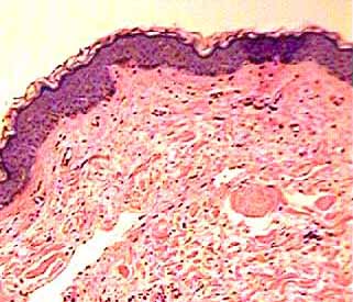
Normal skin is shown at upper left.
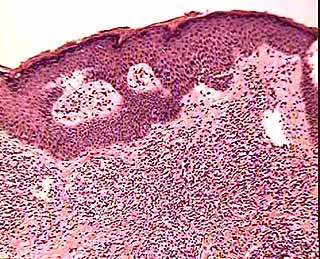
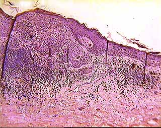
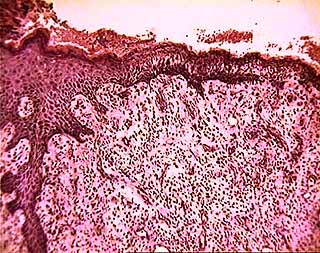
The remaining three images represent pathology. Each of these involves inflammation.In the upper and lower right images, note the profusion of small dark granules in the dermis. These are the nuclei of white blood cells, representing the presence of an inflammatory infiltrate. Read the associated pathology reports for additional description.
|
Normal skin |
Insect bite (pathology report) |
|
Melanoma (pathology report) |
Healing wound (pathology report) |
SURGICAL PATHOLOGY REPORT
Accession No: S-xx-11805
Patient Name: xxxxxxxxxxxxx
Age: 82 years Sex: M Status: Outpatient
GROSS DESCRIPTION
Submitted in formalin as "chest lesion" is a tan white skin tissue measuring 1.3 by 1.3 by 0.2 cm. Centrally located on and covering the majority of the skin tissue is a roughened dark brown area measuring 1.1 cm. in greatest dimension. After inking the margin of excision, the specimen is serially sectioned. The specimen is entirely submitted for histologic study.
HISTOLOGY
The sections are of skin tissue with a malignant melanoma in association with a nevus. The melanoma cells have abundant pigmentation in some areas while in others assume a more spindled shape appearance. The melanoma fills the papillary dermis corresponding to Clark's level II. The tumor at its maximum point measures 1.01 mm. The margins of the shave biopsy on the slide are free of the lesion but not all margins are demonstrated. [Enlarged micrograph]
ANATOMIC HISTORY INQUIRY
Accession No: S-xx-13510
Patient Name: xxxxxxxxxxxxx
Age: 12 years Sex: --- Status: ---
Skin tissue with superficial and deep perivascular infiltrates and focal epidermal change consistent with reaction to an insect bite. [Enlarged micrograph]
With the distribution of the lesions, scabies would be a possibility although there is nothing specific in the biopsy.
SURGICAL PATHOLOGY REPORT
Accession No: S-xx-16787
Patient Name: xxxxxxxxxxxxx
Age: 18 years Sex: M Status: Referral
CLINICAL HISTORY
Growing lesion from antecubital space of the left arm. The lesion had arisen in approximately six weeks and was ulcerated.
GROSS DESCRIPTION
Submitted in formalin is a gray tan, somewhat elliptical, tissue with a polypoid lesion on the skin surface. ... The polypoid lesion is 0.9 by 1.1 by 0.7 cm. in greatest dimensions. The margin of excision will be marked with black ink.
HISTOLOGY
[Specimen] is a nodular lesion composed of granulation tissue characterized by the capillary proliferation in a background of acute and chronic inflammatory cells. The surface is ulerated with fibrinous exudates on the surface. The process is identical to that which is seen in granulation tissue formation in other reparative processes such as wound healing. The lesion presumably arises from a small injury in which the healing process is characterized by exaggerated granulation tissue formation and becomes a so-called "pyogenic granuloma." It is not a neoplastic lesion. [Enlarged micrograph]
Comments and questions: dgking@siu.edu
SIUC / School
of Medicine / Anatomy / David
King
https://histology.siu.edu/intro/inflskin.htm
Last updated: 14 June 2022 / dgk