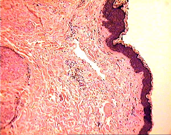
Skin Biopsy Specimen
Magnify a region of this micrograph.
Point and click for more information on a feature.
Features which can be seen in this image include:
- Epithelial tissue of epidermis.
- Connective tissue of dermis.
- Several blood vessels associated with mild perivascular inflammatory infiltrate.
- "Perivascular" simply means "around the blood vessels". "Inflammatory infiltrate" consists of white blood cells which enter the tissue as part of the inflammatory response.
- Although it is not practical to identify reliably most individual cells in this region, the numerous small, roundish, intensely basophilic nuclei seen in this region are characteristic of lymphocytes.
- The inflammatory infiltrate is characterized as "mild" based on the relatively low numbers of white blood cells which appear here. This appearance should be contrasted with a more severe inflammatory infiltrate observable in other slides of inflamed tissue.
- (For more, see inflammation.)
- Smooth muscle.
- In what regions of the body does smooth muscle occur within the skin (besides the tiny arrector pili muscles associated with hair follicles)?
- Many small features (such as individual fibroblasts and collagen fibers) are not individually labelled.
.
Comments and questions: dgking@siu.edu
SIUC / School
of Medicine / Anatomy / David
King
http://www.siumed.edu/~dking2/intro/skinbiop/du1SD3a.htm
Last updated: 11 December 2007 / dgk