Smooth Muscle
muscle
Smooth muscle (unlike skeletal muscle) consists of discrete individual cells (leiomyocytes, a name seldom used). Each smooth muscle cell has own nucleus.
The function of smooth muscle also differs substantially from that of striated muscle.
Neurotransmitter activitation of smooth muscle is fairly diffuse. There are no discrete, well-defined neuromuscular junctions. The motor endings of autonomic axons, where neurotransmitter is released, are not closely associated with individual smooth muscle fibers.
Electrical activitation of smooth muscle is passed from cell to cell by gap junctions.
Smooth muscle of the gut can generate intrinsic rhythmic contraction, independent from direct neural control. Input from the autonomic nervous system increases or decreases the level of this spontaneous activity. (In certain other locations, such as the iris of the eye, neural control is more direct and precise.)
Consult your textbook for more information, including molecular details of contraction and control mechanisms.
Each smooth muscle cell (or "muscle fiber") is just a few microns in diameter but may be two hundred microns long. The nucleus is also elongated, often cigar-shaped.
In histologic sections, the full length of smooth muscle nuclei is only apparent when the plane of section is aligned with the long axis of the cells.
Because the cells that comprise smooth muscle fibers are typically packed closely together, individual cells are usually difficult to resolve, especially at low magnification (as in the thumbnails at right).
In contrast, at the same magnification individual striated muscle fibers are conspicuous as individual units. Each fiber has so many nuclei that any random cross section typically displays at least one and often several, normally located around the periphery of the fiber.
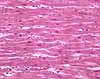
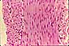
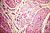
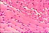
The appearance, or visual texture, of smooth muscle varies dramatically depending on the orientation of the fibers with respect to the plane of section. Note especially the appearance of smooth muscle nuclei.
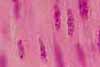
When cut in longitudinal section, smooth muscle nuclei appear very long and sometimes wiggly (due to contraction).
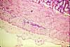
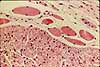
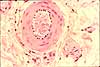
In cross-sectioned smooth muscle, only a fraction of the fibers display nuclei. Recall that each smooth muscle fiber is a single cell, each with its own nucleus. In each such cell, the nucleus occupies only a small proportion of the cell's total length. So nuclei appear only in a similarly small proportion of the fibers cut by any single section.
A very small bundle of smooth muscle can be particularly inconspicuous when cut in cross section. However, one clue is that the cell nuclei appear as small round dots whose apparent size (about 3-4µm) and consistent shape are unlike those of most other cells. (Lymphocytes, the smallest cells that are commonly encountered in connective tissue, have nuclei about 5-6µm in diameter.)
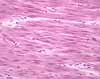
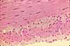
Smooth muscle may also be difficult to recognize when cut obliquely, so that nuclei appear neither small and round nor long and cigar shaped.
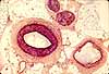
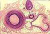
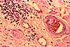
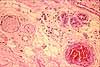
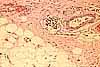
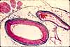
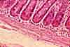
In most histological specimens, some smooth muscle can be found in the walls of arteries and larger veins, as in the examples below.
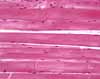
Collagen resembles smooth muscle, in being fibrous and eosinophilic (i.e., pink staining with H&E). However, in most cases collagen can be readily distinguished from smooth muscle by the collagen fibers' irregular size and orientation, and by the haphazard shape and distribution of fibroblast nuclei. (The three thumbnails at right are all from the same tissue sample.)
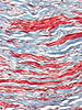
Subtle differences in color can sometimes distinguish smooth muscle from collagen in specimens stained with H&E, but the quality of difference varies from specimen to specimen. To establish this difference on any particular specimen, find a substantial artery and compare its tunica media (muscular layer) with the surrounding adventitial connective tissue. For distinguishing between smooth muscle and collagen, texture (size, shape, orientation of fibers and associated cell nuclei) is usually more useful than color.
However, when it is important to make a quick and reliable distinction between smooth muscle and collagen, a stain that is selective for collagen may be chosen (e.g., one of the trichrome stains).
Examples
 |
 |
 |
 |
 |
 |
 |
 |
|
Comments and questions: dgking@siu.edu
SIUC / School
of Medicine / Anatomy / David
King
https://histology.siu.edu/ssb/smoothm.htm
Last updated: 26 June 2022 / dgk