
Pyramidal Cell of Cerebral Cortex
Historical note: "The year 2006 marks the centenary of the Nobel Prize for Physiology or Medicine awarded to Santiago Ramón y Cajal [1852-1934] and Camillo Golgi [1843-1926]. [Cajal's Nobel Prize lecture (pdf, ~700kB).] We commemorate this centenary with a three-dimensional reconstruction and a quantitative study of a pyramidal cell of a Cajal’s histological preparation. This preparation is one of the 4529 histological preparations personally made by Ramón y Cajal and preserved in the Museum Cajal. The three-dimensional reconstruction of the neuron allows visualizing one important discovery of Ramón y Cajal that constitutes an active field of research in present-day neuroscience: dendritic spines"[from García-López, García-Marín, & Freire (2006) J. Neuroscience 26: 11249-11252].
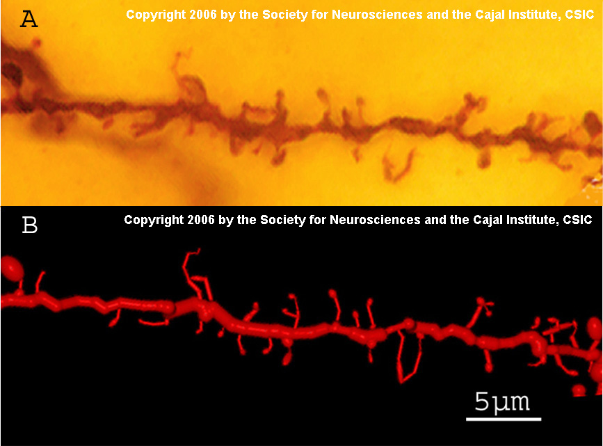
Plasticity (a term introduced by Cajal) of dendritic spine morphology is implicated in memory. Size and shape of dendritic spines influence synaptic strength.
 |
 |
|
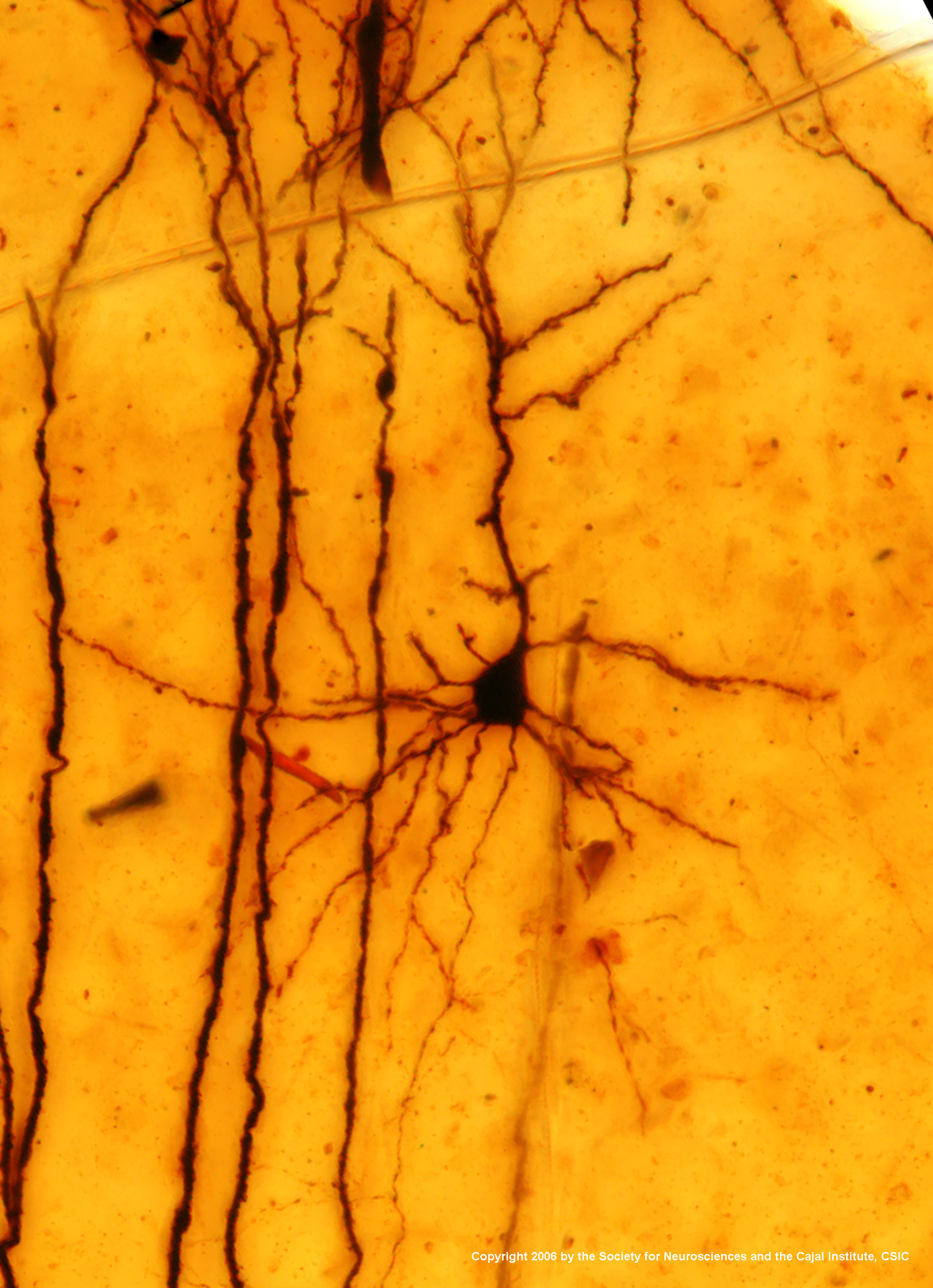
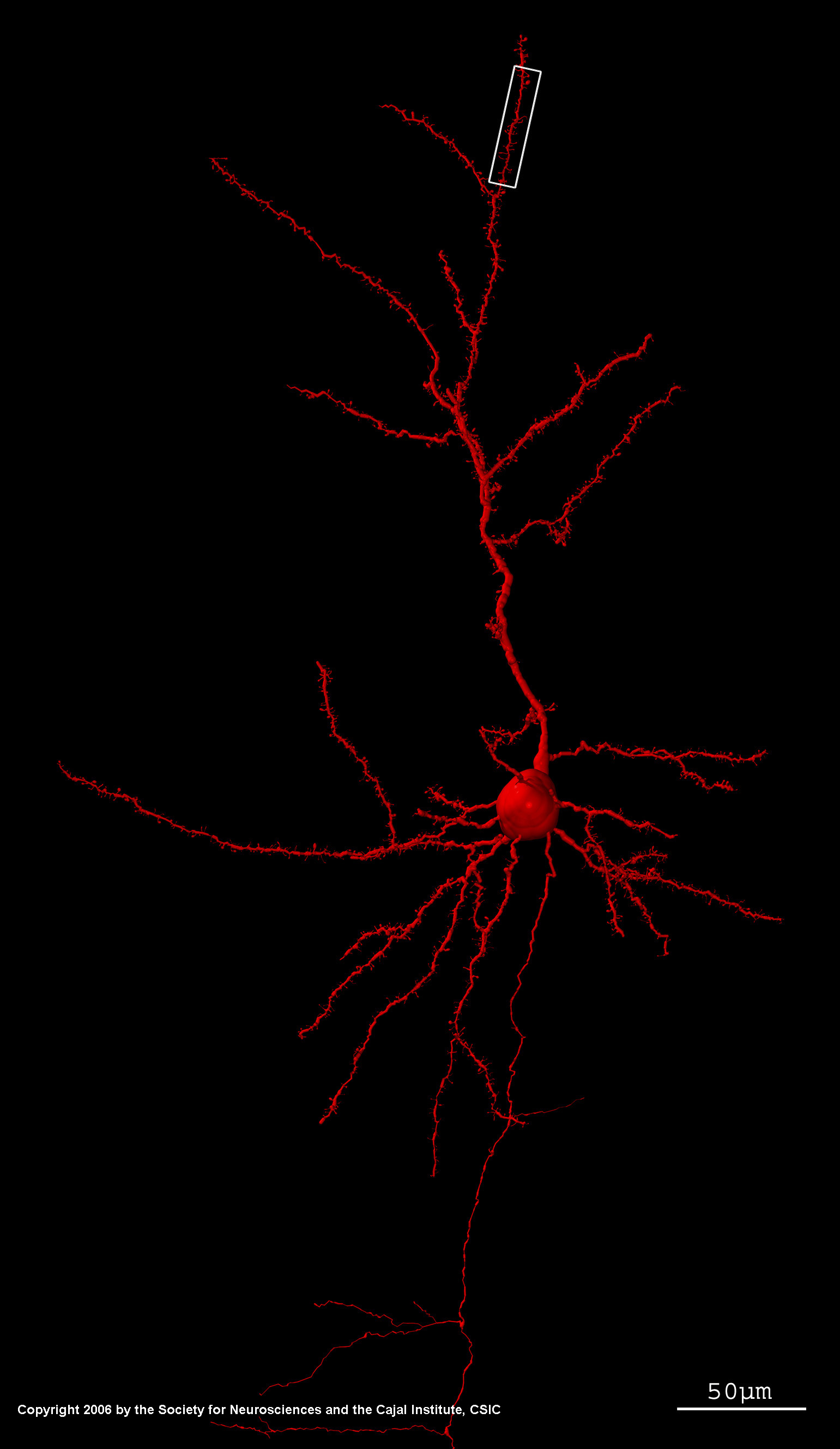
Above: One of Santiago Ramón y Cajal's original preparations shows a pyramidal cell from layer III of mouse cerebral cortex, impregnated with the Golgi method. The long apical dendrite of the cell is quite prominent, extending upward toward the surface of the cerebral cortex. Innumerable other cells (both neurons and glia) remain unstained and are hence invisible. Detail shown below. Right: Computer reconstruction of this neuron. Detail shown below. |
|
|
Images: Copyright 2006 by the Society for Neurosciences and the Cajal Institute, CSIC. Image source: Pablo García-López, Virginia García-Marín, and Miguel Freire (2006) Three-Dimensional Reconstruction and Quantitative Study of a Pyramidal Cell of a Cajal Histological Preparation, Journal of Neuroscience 26: 11249-11252; doi:10.1523/JNEUROSCI.3543-06.2006 |
|
Click on either image above for larger, higher resolution images (1 MByte files).
| Below: Detailed images showing dendritic spines (sites of synaptic communication and memory). |
 |
|
Images: Copyright 2006 by the Society for Neurosciences and the Cajal Institute, CSIC. Image source: Pablo García-López, Virginia García-Marín, and Miguel Freire (2006) Three-Dimensional Reconstruction and Quantitative Study of a Pyramidal Cell of a Cajal Histological Preparation, Journal of Neuroscience 26: 11249-11252; doi:10.1523/JNEUROSCI.3543-06.2006 |
Comments and questions: dgking@siu.edu
SIUC / School
of Medicine / Anatomy / David
King
https://histology.siu.edu/ssb/JNFig1.htm
Last updated: 5 November 2021 / dgk