
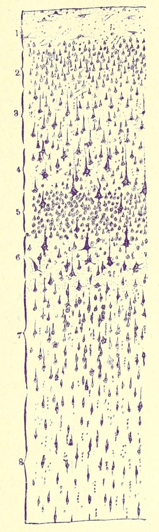
The image below left is a drawing by Cajal, based on a Nissl-stained preparation illustrating cell bodies in the cerebral cortex. Cajal's numbering for cortical layers differs from modern convention. [This image is from Ramón y Cajal's Histologie du Systeme Nerveux de l'Homme et des Vertebres (Maloine, Paris; vol. II, p. 521, 1911).]
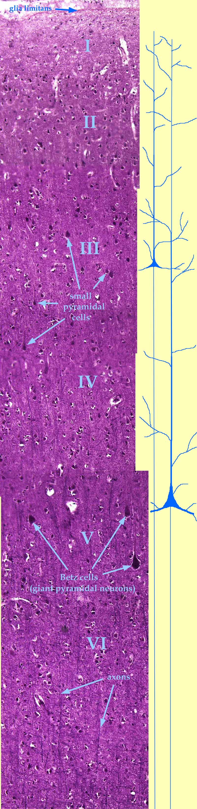
Farther down, this page offers a micrograph across the entire thickness of primary motor cortex (Brodmann's area 4). This is a very tall image. To view it in its entirety, you must scroll down (way down!).
For a lower-magnification overview, click on the thumbnail.
The glia limitans (a continuous layer of astrocyte end feet) is the outermost surface of the brain, immediately beneath the pia mater (which is poorly imaged here).
Designation of the cortical layers I, II, III, IV, V, VI is only approximate. Layers are distinguished by the relative proportions of various cells shapes and fiber distributions; there are no distinct features to serve as "boundaries" for the layers.
The background "feltwork" texture consists of neural processes (dendrites and axons) along with glial processes. This material is sometimes called neuropil.
Some afferent and efferent axons may be visible as thin vertical lines (often in small bundles), especially toward the bottom of the image.
Pyramidal cells may be recognized by their relatively large somata and by their prominent apical dendrites (i.e., the upward "apex" on the "pyramid"). The diagrammatic pyramidal cells alongside the image suggest the extent of these dendrites (although most lateral branches are truncated in the diagram).
So-called "giant pyramidal cells" (also called "Betz cells" after their discoverer, Vladimir Betz, b. 1834) occur only in the primary motor cortex.
Although typically an apical dendrite is conspicuous only near the soma of its pyramidal cell, these are actually very long processes which may extend (with many branches) into the molecular layer (layer I).
Basal dendrites (beginning the lower "corners" of each "pyramid") may extend to either side far beyond the width of this image.
Axons from pyramidal cells project distances which, at this magnification, could equal many tens of meters. Some go to adjacent areas of cortex ("association fibers"). Others cross through the corpus callosum. Those of the upper motor neurons (the giant cells of Betz) cells extend down the pyramidal tract to synapse with spinal motor neurons.
Other neurons and glia cannot be reliably identified at the resolution of this image.
Comments and questions: dgking@siu.edu
SIUC / School
of Medicine / Anatomy / David
King
https://histology.siu.edu/ssb/NM022b.htm
Last updated: 20 January 2026 / dgk