 |
| Image copyright 2008, Tom Deerinck, NCMIR, used with permission. |
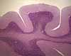
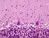
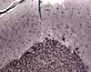
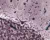
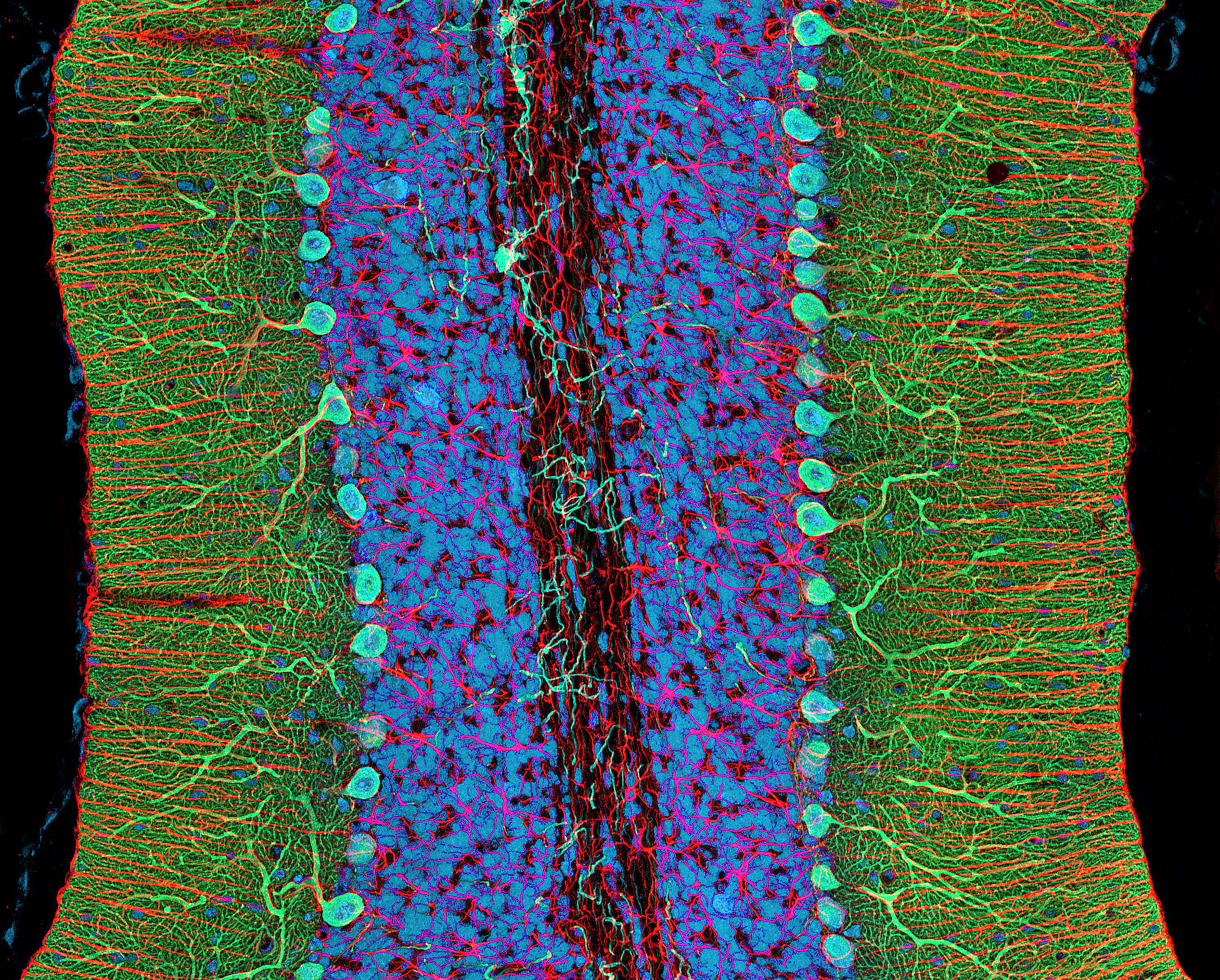
In this "2-photon microscopy" image of cerebellar cortex from the National Center for Microscopy and Imaging Research, fluorescence highlights several structures.
Click here or on the image to open a much larger (2 megabyte) version of this image.
Red color labels GFAP (glial fibrillary acidic protein) in astrocytes. Astrocytes assume their typical shape in the granule cell layer. The radial astrocytic processes in the molecular layer represent Bergmann glia (named after Bergmann, b. 1814), which are important for guiding cell migration during neurogenesis.
Green color labels the IP3 (inositol triphosphate) receptor protein, concentrated in cytoplasm of Purkinje cells (named after Johann Purkinje, b. 1787). Note the extensive ramification of Purkinje cell dendrites in the molecular layer.
Blue color labels DNA, most conspicuously in nuclei of granule cells in the granule cell layer.
Comments and questions: dgking@siu.edu
SIUC / School
of Medicine / Anatomy / David
King
https://histology.siu.edu/ssb/NM041b.htm
Last updated: 18 January 2024 / dgk