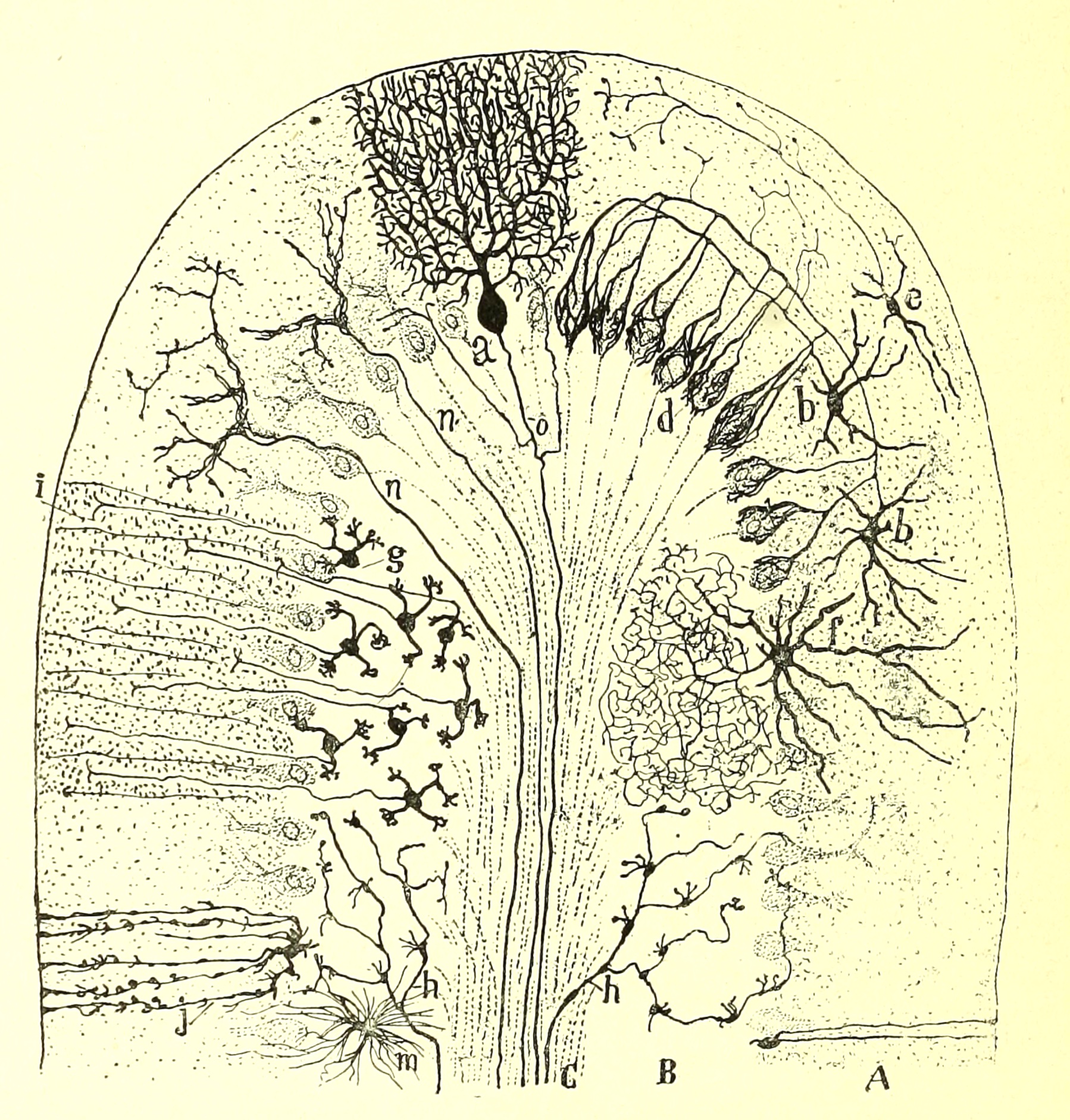

Cerebellar cortex, drawn by Cajal

Image from Ramón y Cajal's Histologie du Systeme Nerveux del'Homme et des Vertebres [accessed at the Wellcome Collection].
Several distinctive nerve cell types characterize the cerebellar cortex; these must be imagined all packed in together, with specific patterns of synaptic connectivity, rather than arrayed into separate regions.
A: molecular layer, B: granular cell layer, C: white matter; a: Purkinje cell, b: basket cells, e: superficial stellate cell, f: large stellate cell (= Golgi cell), g: granule cells, h: mossy fibers, n: climbing fibers.
Comments and questions: dgking@siu.edu
SIUC / School
of Medicine / Anatomy / David
King
https://histology.siu.edu/ssb/cajal-crbllm.htm
Last updated: 19 August 2022 / dgk