

Notes
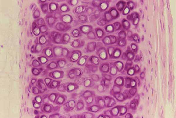
This section of a tracheal cartilage shows the perichondrium on either side. Within the cartilage itself, lacunae are separated by cartilage matrix. Chondrocytes may be seen in a few of the lacunae. Seemingly empty lacunae may represent sites where chondrocytes were inadequately fixed (neither fixatives nor nutrients diffuse quickly through cartilage matrix).
The appearance of cartilage can vary considerably, depending on age and state of development. Because the chondrocytes can secrete matrix but cannot move through the matrix, daughter cells from cell division lie to closer one another than to less closely related cells. This process gives to mature cartilage a characteristic pattern of cell clustering into small groups (not pronounced in this micrograph). Growing cartilage has a lesser proportion of matrix and less conspicuous clustering. Hence this cartilage may come from a still-growing specimen.
Comments and questions: dgking@siu.edu
SIUC / School
of Medicine / Anatomy / David
King
https://histology.siu.edu/ssb/hyalcart.htm
Last updated: 9 August 2021 / dgk