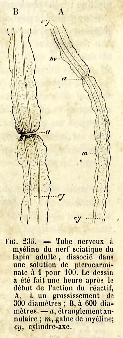
Node of Ranvier, drawing by L. Ranvier
 |
A node of Ranvier appears as a gap in the myelin, where the axon passes from one segment of myelin to the next. This image was taken from an online facsimile of L. Ranvier's Traité technique d'histologie (1875), available at the Wellcome Collection. In 820 pages, this treatise provides an illustrated overview (in French) of histological knowledge in the second half of the nineteenth century. In both drawings here, the node is labelled α. "[Ranvier's] description of nerve fibre nodes was made in a search for how nutrients were continuously exchanged with the blood for nerve cell function... Physiology had demonstrated a loss of motor nerve function by interruption of blood flow and a return to function by perfusion of oxygenated blood... The question was then clear to Ranvier: what is the path for oxygen between oxygenated blood and nerve fibers? For Ranvier, the continuous and impermeable myelin sheath of nerve fibres prevented exchange of fluids and thereby nutrition. He demonstrated the point histologically showing that ... picrocarminate could penetrate fibres, at localized sites identified as interruptions of the myelin sheath..." The above quote is taken from an excellent, illustrated overview of Ranvier's research by Jean Gaël Barbara, available here [from IBRO, the International Brain Research Organization]. |
