
Major blood vessels at base of brain, including the circle of Willis
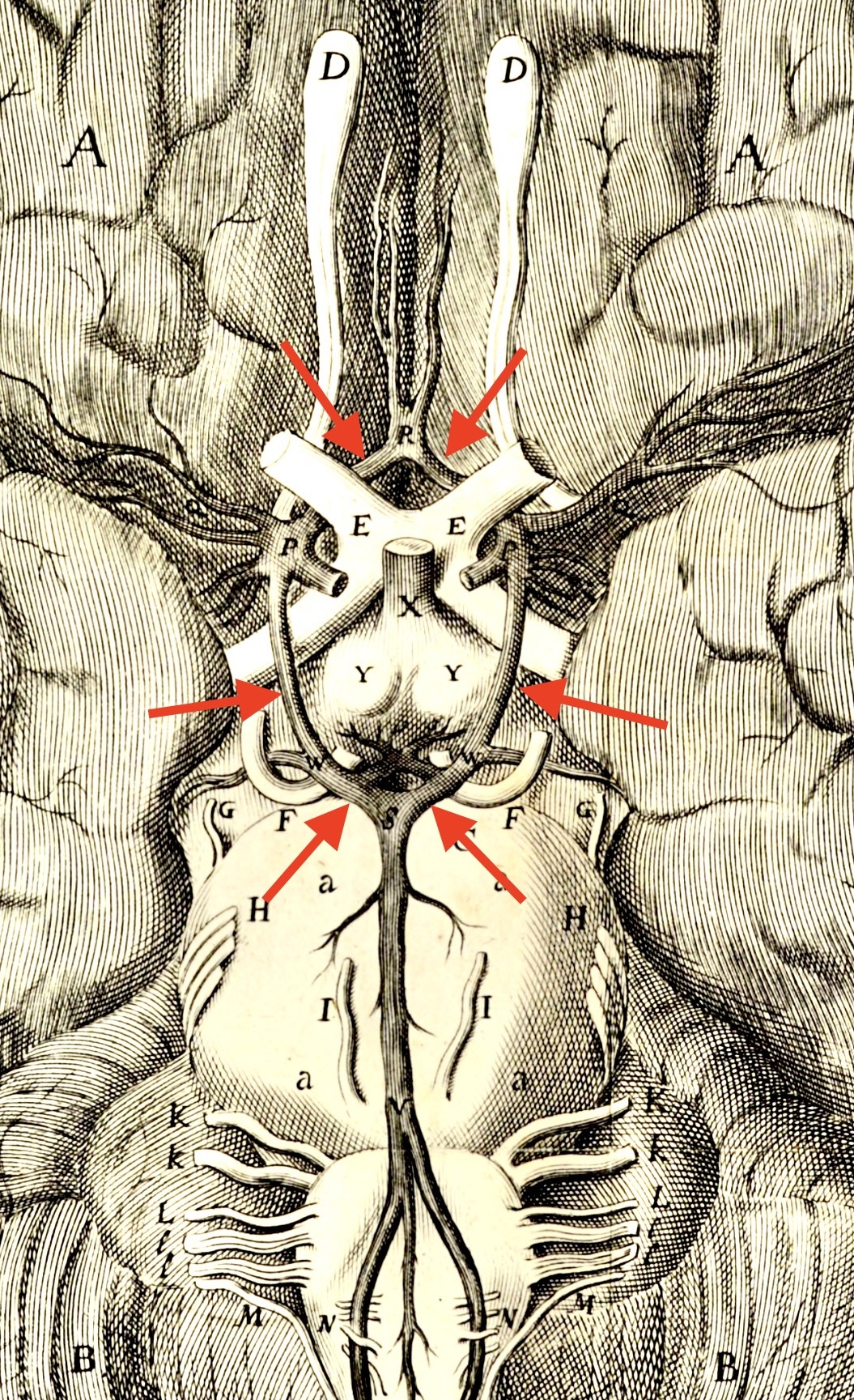 |
Base of the brain, as illustrated in Thomas Willis's Cerebri anatome, 1664. The features shown in this image [adapted from The Wellcome Collection] should appear famiiar to modern students of neuroanatomy. The circle of Willis (red arrows) is a ring of interconnected arteries whose redundancy may help balance blood flow into the brain.
Historical note: The 1600s famously saw the rise of physical and medical sciences. Thomas Willis founded the modern discipline of neurology with his pioneering work, published in 1664: Cerebri anatome: cui accessit nervorum descriptio et usus [Anatomy of the brain, including description and use of nerves]. The images in this volume were drawn by Christopher Wren, the future architect of St. Paul's Cathedral in London. Willis's colleague at Oxford, William Harvey, established the circulation of blood. Willis himself is commemorated in the eponymous circle of Willis. |
Comments and questions: dgking@siu.edu
SIUC / School
of Medicine / Anatomy / David
King
https://histology.siu.edu/ssb/willis-wren.htm
Last updated: 21 May 2025 / dgk