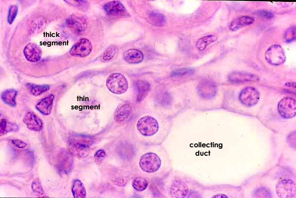


RENAL IMAGE INDEXThis image contrasts three different tubules in the renal medulla:
- A descending thin segment of the loop of Henle, lined by squamous epithelial cells;
- An ascending thick segment of the loop of Henle, lined by cuboidal epithelial cells having eosinophilic cytoplasm and no apparent cell boundaries; and
- A collecting duct, lined by cuboidal epithelial cells having relatively clear cytoplasm and distinct cell boundaries.
(Recall that cuboidal epithelial cells lining tubules typically have round nuclei.
Even the squamous epithelial cells lining loops of Henle usually have round nuclei.)
collecting duct
(not to scale)
Click on the thumbnail at left for lower magnification view.
To compare medullary tubules in longitudinal section, click on one of the thumbnails at right.
Comments and questions: dgking@siu.edu
SIUC / School
of Medicine / Anatomy / David
King
https://histology.siu.edu/crr/RN056b.htm
Last updated: 30 May 2022 / dgk