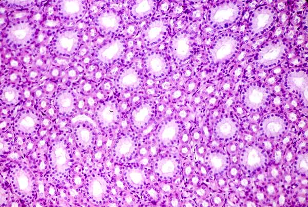


RENAL IMAGE INDEXNote that in this image of the renal medulla, tubules are all cut in cross section, indicating that they are parallel to one another and are arranged perpendicular to the plane of section.
Click on the thumbnail at right to compare with a different plane of section.
The larger tubules in this image are collecting ducts (examples *). The smaller tubules are ascending thick segments of loops of Henle (examples +). Descending thin segments are also present in similar numbers but are inconspicuous at this magnification due to their very thin (squamous) wall.
To view medullary tubules at higher magnification, click on one of the thumbnails below.
Comments and questions: dgking@siu.edu
SIUC / School
of Medicine / Anatomy / David
King
https://histology.siu.edu/crr/RN021b.htm
Last updated: 30 May 2022 / dgk