
| gland | kidney |
 |  |
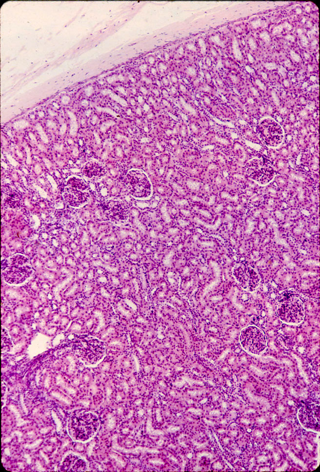
| gland (parotid) | kidney (cortex) |
 |  |
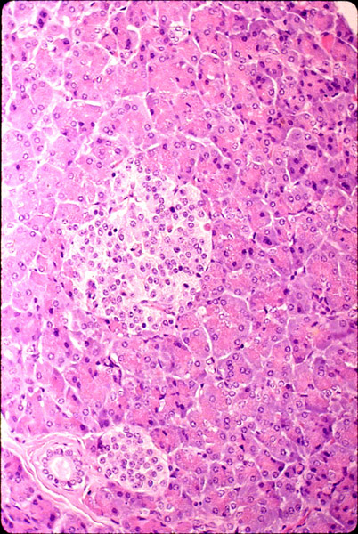
| gland (pancreas) | kidney (cortex) |
 |  |
| gland (pancreas) | kidney (medulla) |
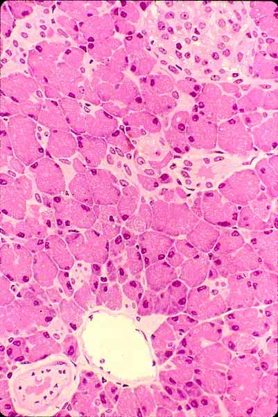 | 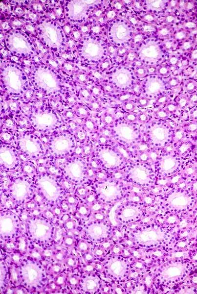 |
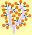
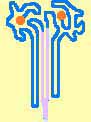
The kidney is built according to the basic structural plan of an exocrine gland such as pancreas or parotid gland (also see common patterns of tissue organization).
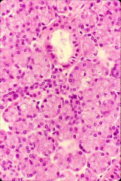
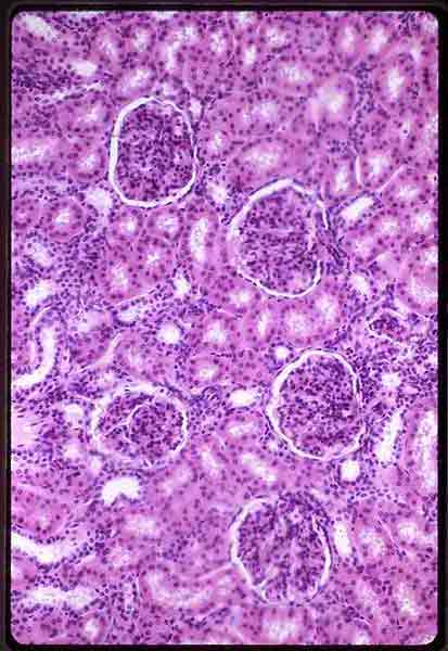
Despite this fundamental similarity, the appearance of kidney differs markedly from that of a typical gland. As can be seen by comparing the image pairs above, whereas a compound acinar gland appears to consists mostly of balls secretory cells (acini) with only occasionally noticeable tubules (ducts), the kidney appears to consist mostly of tubules, with occasional characteristic glomeruli (which are analogous to secretory acini).
Stroma (supporting connective tissue and capillaries) is normally inconspicuous in both kidney and these two glands.
(The pairs of images above are not matched for magnification.)
Return to Kidney Overview
Comments and questions: dgking@siu.edu
SIUC / School
of Medicine / Anatomy / David
King
https://histology.siu.edu/crr/kidney-vs-gland.htm
Last updated: 26 October 2022 / dgk