
Image courtesy of Anthony Huang
Notes
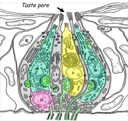
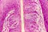
Taste bud functioning remains a topic for research. This diagram illustrates four cell types within a single taste bud, as distinguished by ultrastructural features, patterns of gene expression, and functions. Types I, II, and III taste cells are also classified as:
Glial-like cells (type I, gray in diagram).
Receptor cells (type II, yellow in diagram, associated with nerve endings but without classically-recognizable synapses).
Presynaptic cells (type III, blue in diagram).Colored pink in the diagram are basal cells (B).
Green color indicates nerve fibers.
Non-sensory cells of the tongue's stratified squamous epithelium (E) are not colored in this diagram.
For more on tastebuds, see Chaudhari & Roper (2010) Journal of Cell Biology 190:285-296, doi: 10.1083/jcb.201003144
Related examples:
 |
 |
 |
 |
Comments and questions: dgking@siu.edu
SIUC / School
of Medicine / Anatomy / David
King
https://histology.siu.edu/erg/Huang-tastebud.htm
Last updated: 31 August 2021 / dgk