Oral Cavity
The oral cavity is specialized for sensory discrimination (taste),
mechanical processing (chewing), initial lubrication and enzymatic
digestion (salivary secretion), and immune
surveillance (tonsils), with a protective stratified
squamous epithelium throughout.
Clinically, the oral cavity provides an easy opportunity for a revealing examination
of a mucosal surface (e.g., "Please open your mouth and say, 'AH' ").
For an extreme example (candidiasis), see WebPath
(gross image) and WebPath
(micrograph).
 The
oral mucosa varies from site to site within the oral cavity, but everywhere
the epithelium is protective stratified
squamous. This epithelium is
partially keratinized on gums and hard palate
and on filiform papillae of tongue.
It is non-keratinized elsewhere. Lamina
propria is unspecialized. A muscularis mucosae
is not present. Deep to the epithelial surface, minor
salivary glands
are common.
 Tongue
The mucosal surface of the
tongue, the part you can see when the patient says "AH," displays
several specialized epithelial variations, including papillae of various
shapes. Variations in gross appearance can be clinically revealing.
(For an extreme example, see WebPath
(gross image) and WebPath
(micrograph).).
In addition to its clinical value as a readily-observed
"window" onto a mucosal surface, the tongue also provides excellent
opportunities for learning histology, with examples of all the basic tissue
types appearing in a variety forms.

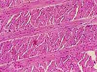
 The bulk of the tongue consists of striated muscle
fibers arranged in bundles along three mutually perpendicular axes, so
any plane section is likely to reveal fibers cut both transversely and longitudinally.
Bundles of myelinated nerve fibers
are usually easy to find within the muscle of the tongue.
 The surface of the tongue is covered by stratified
squamous epithelium, modified on the upper surface into filiform
papillae. These papillae comprise the whitish "fuzz"
over most of the lingual surface. They have keratinized tips (hence their whitish
color) and provide roughness which contributes to the tongue's food-handling
ability. (The name filiform means "file-like." The resemblance
to a file is obvious if you've ever felt a cat's tongue, whose filiform
papillae heavily keratinized.)

 Scattered among the filiform papillae are occasional fungiform papilla. Taste
buds are found on fungiform papillae. Far back on the tongue are a few relatively large
circumvallate papillae.
The thumbnail at right shows foliate (leaf-shaped) papillae
from rabbit tongue, which in section appear similar to human fungiform
papillae.
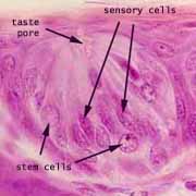
 Taste buds are oval clusters of elongated cells which extend across the thickness
of the epithelium, from the lamina propria
to the taste pore at the surface.
 Within a taste bud, each sensory cell has microvilli
in the taste pore at its apical end. These allow contact with the external
medium. At its basal end, each sensory cell makes synaptic contact with
fibers of the facial nerve (CN VII) or glossopharyngeal nerve
(CN IX).
Tonsils
Tonsils are lymphoid structures
located in the mucosa of the tongue, palate, and pharynx which provide sites
where immune surveillance cells (lymphocytes)
can encounter foreign antigens which enter the body through the mouth or nose. This region is sometimes called
Waldeyer's ring (commemorating Wilhelm Waldeyer, b. 1836)
 Each tonsil consists of an epithelial crypt (invaginated pocket) surrounded by
dense clusters of lymph nodules, each with a germinal center where lymphocytes
proliferate. The nodules are embedded in a mass of diffuse lymphoid
tissue that consists of lymphocytes migrating to and from the germinal centers.
 The epithelium lining a crypt corresponds with that on the adjacent surface
-- stratified squamous in the tongue and palate, or pseudostratified
columnar in the pharynx. In either case, the epithelium may be heavily
infiltrated with lymphocytes, and
the crypt may be filled with lymphocytes and other debris.
The lymphoid tissue of the tonsils is
similar to that of Peyer's patches and appendix.
These structures, together with other more diffuse lymphoid tissue, constitute
the Gut-Associated Lymphoid Tissues, or GALT.
For more on GALT (or, more generally, MALT for Mucosa-Associated
Lymphoid Tissues), consult your histology text (e.g. pp. 134-5 in Stevens
& Lowe).
Palate
 The hard palate has a partially-keratinized epithelium, much of which is
firmly attached by a fibrous submucosa to underlying bone.
The soft palate has a non-keratinized epithelium, with underlying
minor salivary glands and striated
muscle. The largest tonsils (the palatine
tonsils) are embedded in the sides of the soft palate.
Oral cavity examples:
Comments and questions: dgking@siu.edu
SIUC / School
of Medicine / Anatomy / David
King
https://histology.siu.edu/erg/oralcav.htm
Last updated: 16 May 2023 / dgk
 The
oral mucosa varies from site to site within the oral cavity, but everywhere
the epithelium is protective stratified
squamous. This epithelium is
partially keratinized on gums and hard palate
and on filiform papillae of tongue.
It is non-keratinized elsewhere. Lamina
propria is unspecialized. A muscularis mucosae
is not present. Deep to the epithelial surface, minor
salivary glands
are common.


 The bulk of the tongue consists of striated muscle
fibers arranged in bundles along three mutually perpendicular axes, so
any plane section is likely to reveal fibers cut both transversely and longitudinally.
Bundles of myelinated nerve fibers
are usually easy to find within the muscle of the tongue.


