
Placenta and Umbilical Cord

The placenta might be helpfully understood as a "parasite" feeding on blood from the endometrium. Imagine scooping out a portion of the endometrium. The resulting bowl will fill with blood from broken vessels in the endometrial stroma. Now lay a cover over the bowl, and imagine many "roots" extending down from the cover into the blood-filled hollow. The roots can can absorb oxygen and nutrients from the blood in which they are bathed.
The cover is the chorionic plate of the placenta. The "roots" are the chorionic villi.
Both the placenta and the chorionic villi (orange in the cartoon diagram to right) are entirely fetal tissue. "Anchoring villi" attach the placenta to maternal tissue of the endometrium (green in the diagram). Smaller branching villi extend out into the intervillous space. Fetal circulation (red and blue in the diagram) passes down the umbilical cord, though vessels in the villi, and back up the umbilical cord.
Maternal blood (pink in the diagram) "spills" from open endometrial arteries (the spiral arteries) into the intervillous space (pink in the diagram), and returns into endometrial veins. Thus the chorionic villi are surrounded and bathed by "lakes" of maternal blood. Within the intervillous space, maternal blood is not contained by blood vessels.
The surface of the chorionic villi is an epithelial layer, the fetal syncytiotrophoblast, which has the ability to grow invasively into the maternal endometrium. The syncytiotrophoblast also has microvilli on the surface for absorbing nutrients from maternal blood.
In the extravagant name "syncytiotrophoblast," syncytio- tells us that this is a syncytium, a tissue in which many nuclei occupy a large cytoplasmic volume without separation into individual cells (skeletal muscle fibers are a more familar example of syncytial tissue); tropho- tells us that this tissue provides nutrition; and -blast tells us that it is growing.
Beneath the syncytiotrophoblast (i.e., toward the core of the villus), is the cytotrophoblast, also called the layer of Langhans (commemorating Theodor Langhans, b. 1839). This is a layer of cuboidal cells which eventually disappear. (The cytotrophoblast also forms trophoblast columns, masses of cells filling the ends of anchoring villi.) Maternal endometrial stromal tissue adjacent to the placenta differentiates into large decidual cells (so named because the outer layer of the endometrium is shed at birth along with the placenta). Decidual cells may intermix with fetal cells in the cytotrophoblast. The boundary between maternal and fetal tissue is immunologically interesting.
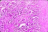
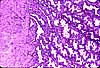
For micrographs of placenta, see WebPath (decidual cells), WebPath (first trimester chorionic villi), WebPath (second trimester chorionic villi), and WebPath (third trimester chorionic villi).
Forming the core of each chorionic villus is mesenchymal stroma containing fetal blood vessels.
- Fetal circulation is entirely closed, confined to vessels within the chorionic villi.
- Maternal blood flow though the placenta is open. "Lakes" of maternal blood fill the intervillous space, uncontained by any endothelial lining.
Between maternal blood and fetal blood lies the thin syncytiotrophoblast and the fetal capillary endothelium, across which all exchange of gases, nutrients, hormones, and wastes occurs.
Perhaps surprisingly, this boundary between fetal and maternal tissue allows some transfer of cells between mother and fetus during pregnancy. The outcome can be a cellular chimera.
☞ As a result of such cellular transfer, many women's bodies include cells with the genotype of a fetus from a previous pregnancy, while the bodies of many individuals include some cells with the genotype of their mother. This in turn has some potentially significant genetic and medical consequences. For further explanation, see microchimerism. For an extended, readable discussion by a celebrated science reporter, see Chapter 13, "Chimeras," in Carl Zimmer's 2018 book about heredity, She Has Her Mother's Laugh.
The umbilical cord is basically just a conduit carrying fetal blood to and from the placenta. It normally contains two arteries and one vein, surrounded by extensive mesenchymal tissue ("Wharton's jelly"). Umbilical cord tissue can be utilized as a source for embryonic stem cells.
 |
 |
|
|
SIUC / School
of Medicine / Anatomy / David
King
https://histology.siu.edu/erg/placenta.htm
Last updated: 14 June 2025 / dgk