
Notes
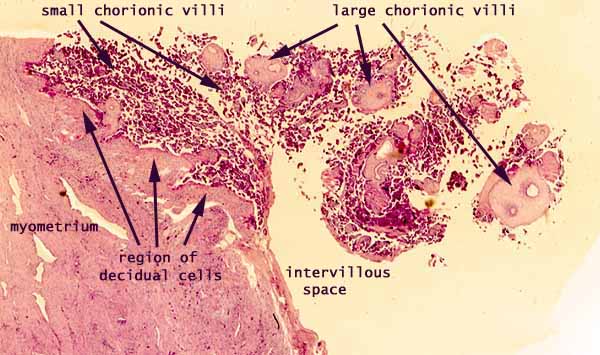
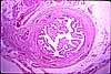
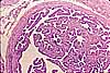
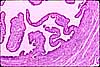
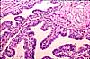
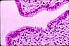
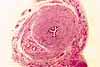
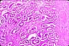
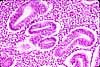
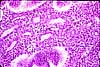
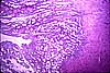
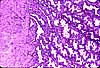
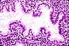
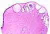
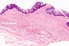
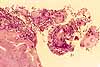
In this image, many chorionic villi are seen in the vicinity of the uterine wall. Both large and small chorionic villi have the same basic structure, a surface of syncytiotrophoblast surrounding a core of mesenchyme containing fetal blood vessels. Fetal blood vessels are visible here only in the largest villi. The small villi vastly increase the surface area available for material interchange with maternal blood in the intervillous space. Decidual cells are large cuboidal cells derived from endometrial stroma.
(For additional micrographs, see WebPath (decidual cells), WebPath (first trimester chorionic villi), WebPath (second trimester chorionic villi), and WebPath (third trimester chorionic villi).)
Related images:
 |
 |
|||
 |
 |
 |
 |
 |
 |
 |
 |
 |
 |
 |
 |
 |
 |
 |
Comments and questions: dgking@siu.edu
SIUC / School
of Medicine / Anatomy / David
King
https://histology.siu.edu/erg/RE051b.htm
Last updated: 20 May 2022 / dgk