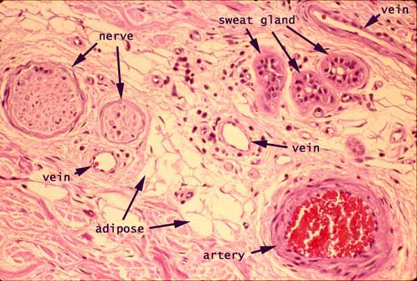

Skin, dermis

Notes
A small amount of subcutaneous adipose tissue appears near the center of this image. Fibrous connective tissue of the dermis occupies the remaining regions and includes a conspicuous artery, several small veins, a portion of a sweat gland, and a pair of small peripheral nerves.
The pink color in this image represents eosinophilic material, in this case mostly one of the following:
- Most of the pink material represents extracellular collagen fibers.
- Color alone does not serve to distinguish the nerve from surrounding collagen; this distinction is made visually on the basis of texture (with the nerve being circumscribed by its surrounding epineurium).
- The somewhat deeper pink encircling the artery represents the cytoplasm of smooth muscle cells.
The pale background color represents unstained material, in this case one of the following:.
- Near the center, the open spaces represent the lipid droplets within adipocytes. (Intact adipocytes appear round, with continuous margins. Adipocytes which have been damaged during specimen preparation appear as irregular shapes with broken margins.)
- Within the veins and the artery, the pale space represents blood plasma (or, more accurately, the lumen where blood would flow in living tissue).
- Within the sweat gland, the tiny, centrally-located clear space represents the glandular lumen, continuous with the outside world.
- Elsewhere, the thin interconnected pale spaces between collagen fibers represent connective tissue ground substance.
The very dark purple (nearly black) spots are cell nuclei. At this resolution, they cannot be individually identified except by context.
- Nuclei adjacent to the lumen of a blood vessel belong to vascular endothelial cells.
- Nuclei in the muscular wall of an artery may belong to smooth muscle cells.
- Irregular nuclei scattered at random among collagen fibers belong mostly to fibroblasts. Some may also belong to macrophages, mast cells, or capillary endothelial cells.
- Thin nuclei adjacent to large lipid droplets may belong to adipocytes or to fibroblasts or to capillary endothelium located between the adipocytes.
Bright red within the artery represents clumped-together masses of red blood cells.
Comments and questions: dgking@siu.edu
SIUC / School
of Medicine / Anatomy / David
King
https://histology.siu.edu/intro/IN010b.htm
Last updated: 11 June 2022 / dgk