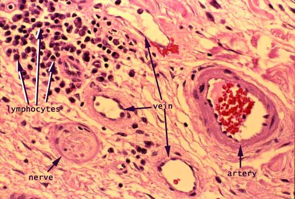

Skin, dermis

Notes
Fibrous connective tissue of the dermis occupies most of the area of this image. Included are a conspicuous artery, several small veins, a small peripheral nerve, and a cluster of lymphocytes representing mild perivascular inflammatory infiltrate.
The pink color in this image represents eosinophilic material, in this specimen mostly one of the following:
- Most of the pink material represents extracellular collagen fibers.
- Color alone does not serve to distinguish the nerve from surrounding collagen; this distinction is made visually on the basis of texture (with the nerve being circumscribed by its surrounding epineurium).
- The somewhat deeper pink encircling the artery represents the cytoplasm of smooth muscle cells.
The pale background color represents unstained material, in this specimen one of the following:
- Within the veins and the artery, the pale space represents blood plasma (or, more accurately, the lumen where blood would flow in living tissue).
- Elsewhere, the thin interconnected pale spaces between collagen fibers represent connective tissue ground substance.
The very dark purple (nearly black) spots are cell nuclei. At this resolution, they cannot be individually identified except by context.
- Nuclei adjacent to the lumen of a blood vessel belong to vascular endothelial cells.
- Nuclei in the muscular wall of an artery may belong to smooth muscle cells.
- Irregular nuclei scattered at random among collagen fibers belong mostly to fibroblasts. Some may also belong to macrophages, mast cells, or capillary endothelial cells.
Bright red within the artery represents many red blood cells.
Comments and questions: dgking@siu.edu
SIUC / School
of Medicine / Anatomy / David
King
https://histology.siu.edu/intro/IN011b.htm
Last updated: 9 June 2022 / dgk