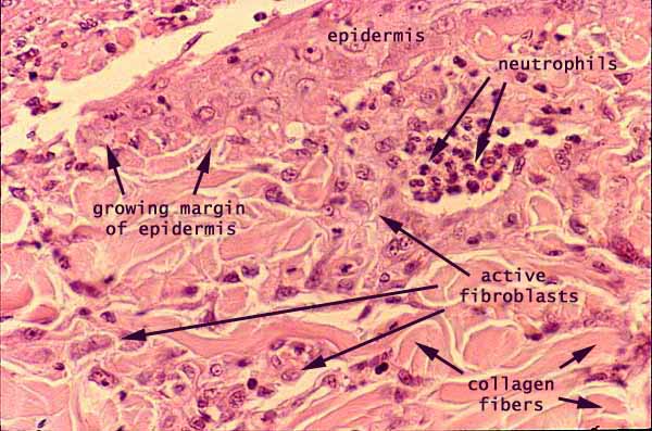

Skin, healing injury.

Epidermis is growing down from top center toward the left, in this image of skin healing after a minor injury.

Context for this image may be better appreciated at lower magnification. Click here or on one of the thumbnails at right to view this specimen at lower magnification.
The epithelial quality of the newly-formed epidermis is apparent, even though well-developed layers have not yet developed.
In fibroblasts that have become active, cytoplasm is more evident and nuclei are more euchromatic than in resting fibroblasts encountered in more typical skin preparations. Also note that the collagen fibers in this region of scar formation are larger than typical for papillary dermis.
The cluster of inflammatory cells (neutrophils) reflects a recent history of inflammation triggered by the injury and possible contamination.
Comments and questions: dgking@siu.edu
SIUC / School
of Medicine / Anatomy / David
King
https://histology.siu.edu/intro/IN021b.htm
Last updated: 12 June 2022 / dgk