
Epithelium and Gland,
Frog Skin
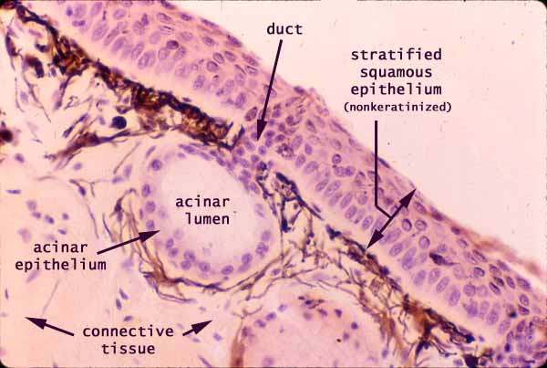
This image shows the basic epithelial configuration of a gland, with surface epithelium (here a nonkeratinized, stratified squamous epithelium) invaginating to form a simple gland, with one single secretory unit connected to the surface by a short duct.
In this specimen, the secretory unit takes the form of a round ball of cells, called an acinus.
Secretory cells may also be organized into elongated tubules rather than rounded acini (as in the sweat gland in the thumbnail at right).
Both the duct and the secretory portion of the gland are formed from cuboidal epithelium, with round nuclei centrally placed within boxy cells.
Most exocrine glands have this same basic epithelial configuration of secretory unit and duct. However, larger compound glands (such as salivary glands or pancreas) have many acini connecting to the surface by a branching duct tree.
The brown / black material in this specimen of frog skin is the pigment melanin, within melanocytes.
Comments and questions: dgking@siu.edu
SIUC / School
of Medicine / Anatomy / David
King
https://histology.siu.edu/intro/IN031b.htm
Last updated: 7 August 2021 / dgk