

Notes
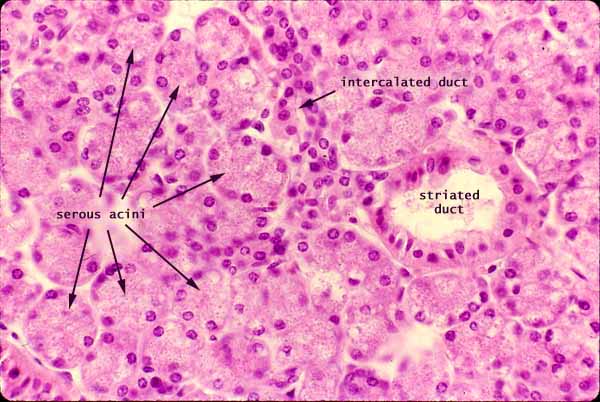
The parotid salivary gland is a compound, acinar, serous gland. Unlike all other salivary glands, the parotid gland includes no mucous cells.

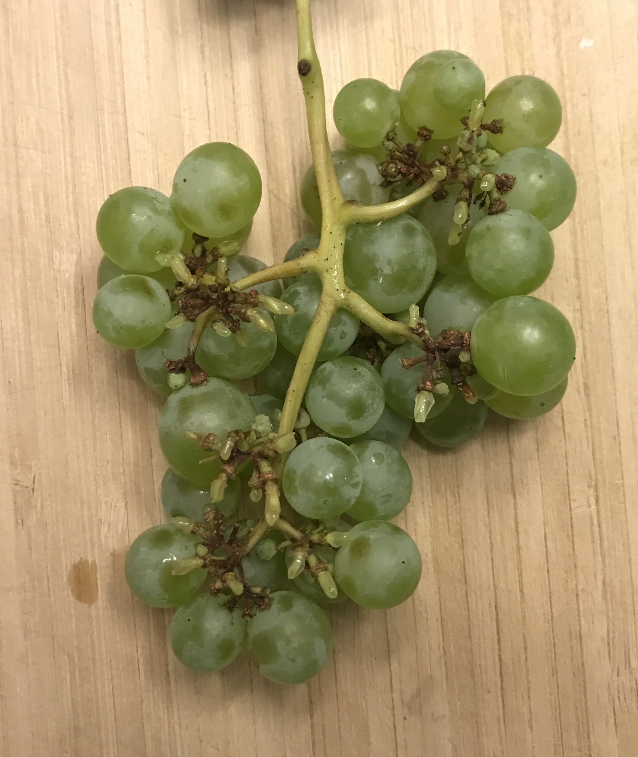
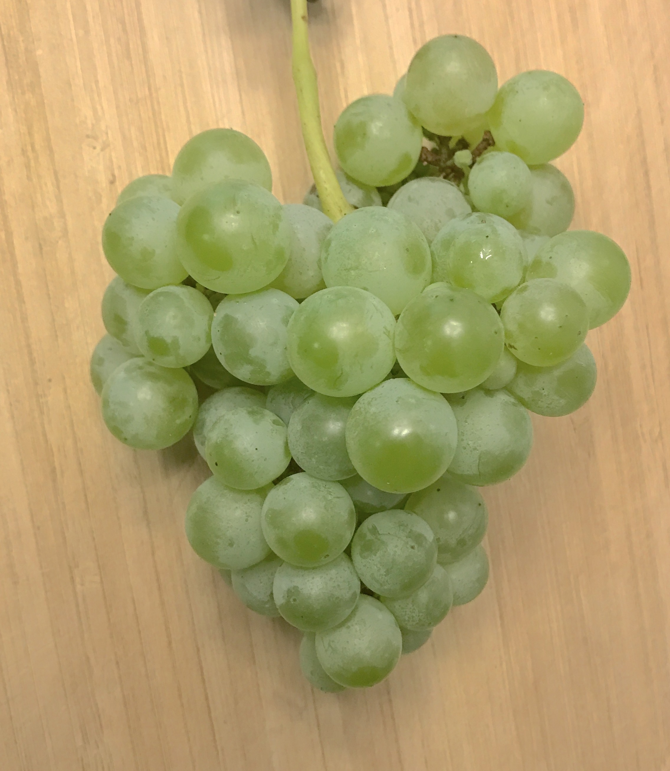
 "Compound acinar" glands have their clusters of secretory cells and ducts configured like grapes and stems in a bunch of grapes.
In the micrograph above, most of the specimen appears packed with secretory acini. These acini are cut in random planes, so that most look like solid lumps made of cells having various sizes and shapes. The acinar lumen is visible only when the acinus is sliced neatly across the middle, as in the thumbnail at right. In such a slice, the cells look like slices of pie, with the lumen in the center.
Individual acini are drained by small intercalated ducts. These in turn drain into striated (or "secretory") ducts, whose cells are specialized for concentrating the secretory product. Cells lining the striated duct pump water and ions across the epithelium, from the duct lumen and into interstitial fluid.
Glandular stroma is not particularly noticeable in this image. Nevertheless, whether visible or not, each acinus is surrounded by a thin envelope of capillaries and connective tissue. (In the "bunch of grapes" analogy, stroma occupies the spaces between the grapes.) Although not seen in this image, adipocytes are also common in the parotid gland.

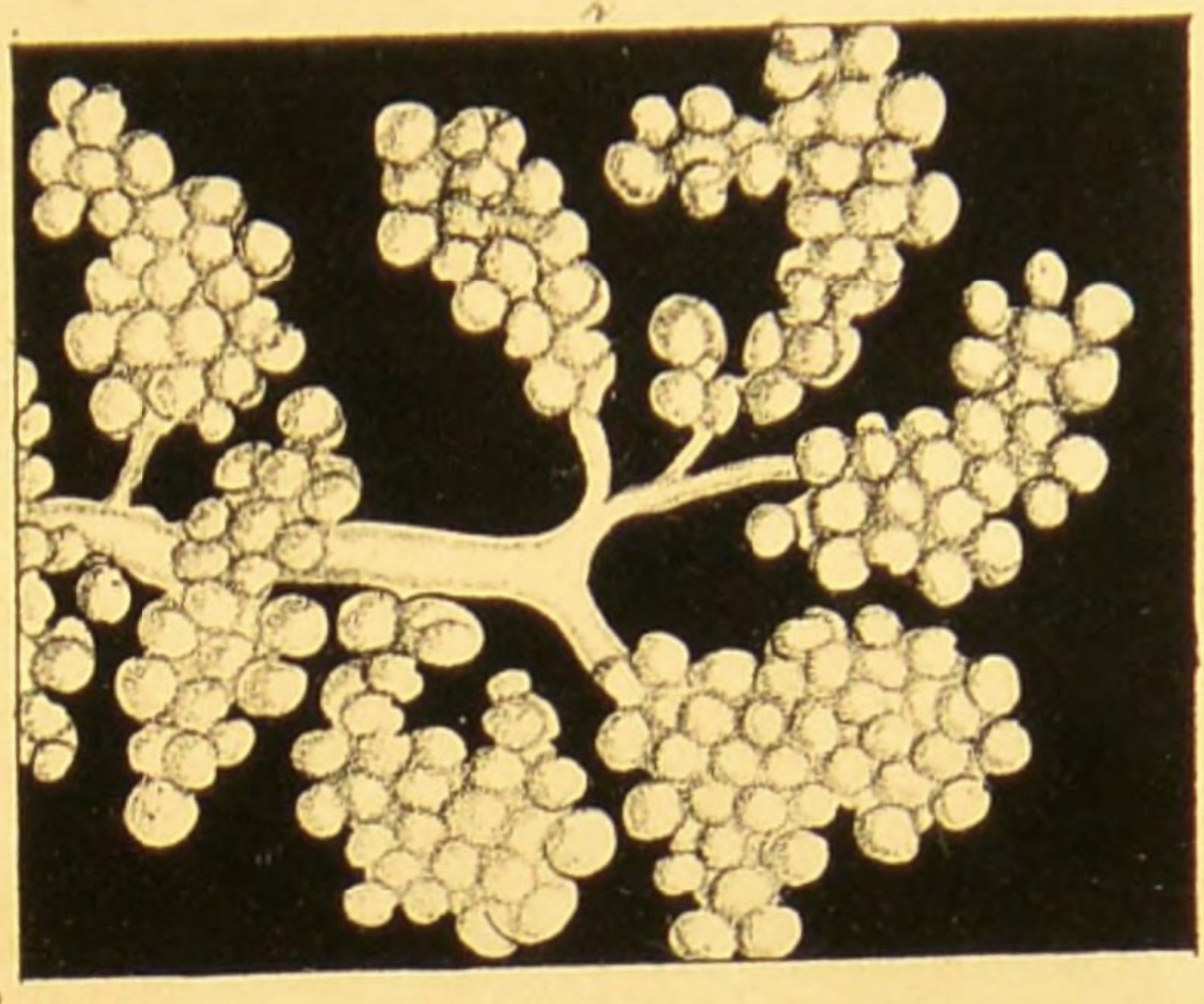
Historical note: The drawing at left, of human parotid gland, comes from the first English-language textbook of microscopic anatomy, by Arthur Hill Hassall, 1849. (Imaged accessed at the Welcome Collection.)
Related examples:
 |
 |
 |
||
 |
 |
 |
 |
|
 |
 |
 |
 |
 |
 |
 |
 |
Comments and questions: dgking@siu.edu
SIUC / School
of Medicine / Anatomy / David
King
https://histology.siu.edu/erg/GI121b.htm
Last updated: 24 January 2023 / dgk