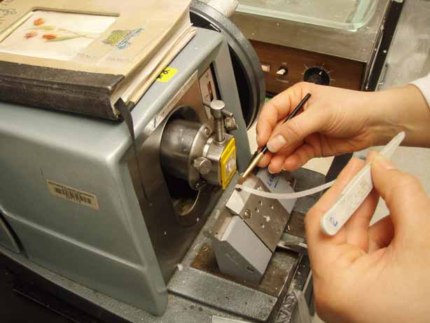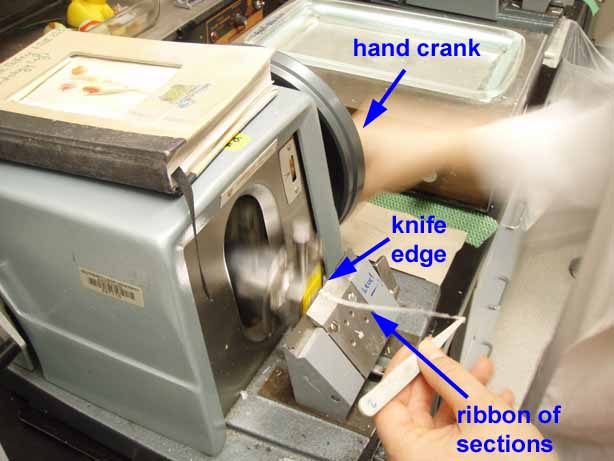
A microtome is used for sectioning -- cutting very thin slices (sections) from a tissue sample.
Historical note: Some sources attribute the development of the first practical microtome to Jan Purkinje (b. 1787), after whom Purkinje cells of cerebellum and Purkinje fibers of heart are named.
- A microtome has the essential machinery of a baloney-slicer. Essential elements include:
- a cutting edge; this may be a razor, a heavy knife, or (for electron microscopy) a piece of broken glass or a finely sharpened gem-quality diamond.
- a specimen holder.
- a screw to advance the specimen toward the blade (ultramicrotomes
may use carefully-controlled thermal expansion in lieu of a screw);
this machinery is typically hidden within a cover. - a crank, such that each turn of the crank raises the specimen, advances it, and then lowers it across the blade.
- Sections are typically floated on a water bath before transfer to a slide.
- A nice video clip of this process is included in an online article in the New York Times, published 9/9/2025 in the context of diagnosing C.T.E. (chronic traumatic encephalopathy).

