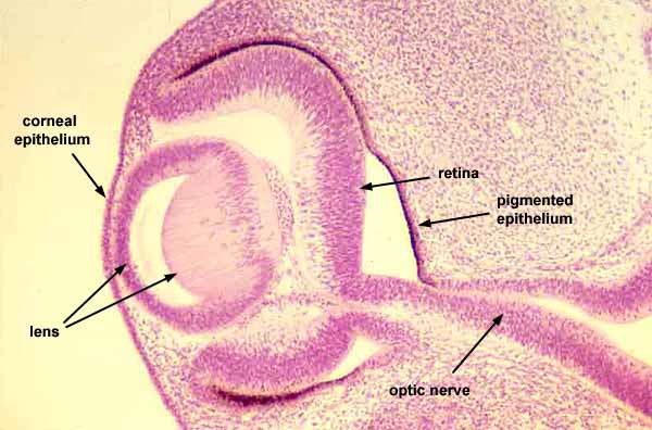

Embryonic eye: lens vesicle and optic cup

SSB IMAGE INDEX

For explanation, see eye embryology.
To view this region in context, click on thumbnail at left.
For views of mature lens and retina, click on thumbnails at right.
Where the embryonic layers have not yet finished coming together, this image shows separation between neural retina (here labelled simply "retina") and pigmented epithelium. Apposition between these two layers remains inherently fragile and forms a site where retinal detachment can occur.
Comments and questions: dgking@siu.edu
SIUC / School
of Medicine / Anatomy / David
King
https://histology.siu.edu/ssb/EE016b.htm
Last updated: 1 August 2023 / dgk