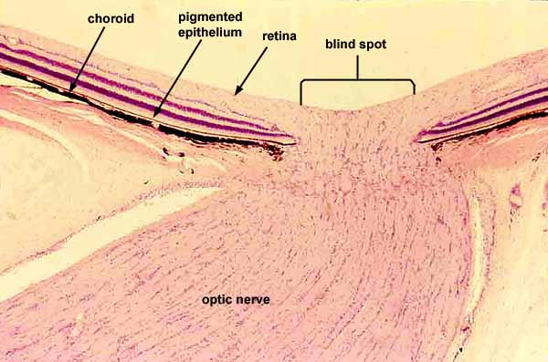

Eye: retina, choroid, and optic nerve

SSB IMAGE INDEXThe retina includes all the tissue layers from the top surface (where the "retina" arrow points) down to the pigmented epithelium.
During normal vision, light would come down from the top of this image and pass through most of the retinal layers before reaching the photoreceptor layer, the pale layer just above the thin brown layer of pigmented epithelium.
The innermost layer of the retina (uppermost in this image) consists of nerve fibers that travel along the surface of the retina before leaving the eyeball through the optic nerve.
A "blind spot" results where these optic nerve fibers penetrate the retina, leaving a small area without photoreceptor cells where no image information is collected.
The choroid, a heavily pigmented layer of loose connective tissue (black in this image), lies below (external to) all these retinal layers.
External to the choroid lies the sclera, the tough, collagenous "white" of the eyeball (unlabelled on this image).
For higher magnification of the retinal layers , click on the thumbnail at right:
Comments and questions: dgking@siu.edu
SIUC / School
of Medicine / Anatomy / David
King
https://histology.siu.edu/ssb/EE019b.htm
Last updated: 1 August 2023 / dgk