

Historical note: In 1847, William Bowman produced an image of retinal layers that was reasonably accurate for his time.
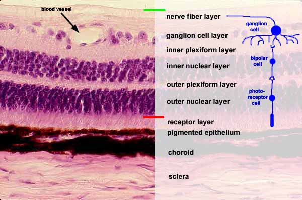
SSB IMAGE INDEXDuring normal vision, light would come down from the top of this image, passing through most of the retinal layers before reaching the photoreceptor layer just above the thin pigmented epithelium.
Note that "inner" and "outer" in the names of layers refer to the eyeball, not to the surface of the retina. Thus "inner" is upward in this image, while "outer" is downward.
- The location of the internal limiting membrane is indicated by the green line at the top of the image (above the nerve fiber layer). This layer is a basal lamina separating nervous tissue of the retina from connective tissue of the vitreous humor.
The layer of nerve fibers contains axons from ganglion cells which travel across the retina to the optic nerve and hence past the optic chiasm into the optic tract and into lateral geniculate nucleus of the thalamus.
- The ganglion cell layer contains the cell bodies of ganglion cells, the cells whose axons project to the brain.
- The inner plexiform layer contains dendrites of ganglion cells synapsing with axons of bipolar cells.
- The inner nuclear layer contains the cell bodies of bipolar cells. Although additional cell types are also present in this layer, most of the nuclei belong to bipolar cells.
- The outer plexiform layer contains dendrites of bipolar cells synapsing with axons of photoreceptor cells.
- The outer nuclear layer contains the cell bodies of receptor cells. Although additional cell types are also present in t his layer, most of the nuclei belong to photoreceptor cells
- The receptor layer contains the photoreceptive outer segments (rods and cones) of receptor cells.
- The pigmented epithelium absorbs light not captured by photoreceptors, and also contributes to the maintenance of rod and cone outer segments.
- The location of the external limiting membrane (composed of processes of Müller glial cells) is indicated by the red line just above the receptor layer. Outer segments of the receptor cells (rods and cones) penetrate this membrane to contact the pigmented epithelium.
NOTE: Thickness of retinal layers and the number of nuclei in each layer varies with location across the retinal surface. To see how the concentration of cells and the resulting thickness of the retina decrease toward the periphery (i.e., with increasing distance from the macula), click here or on thumbnails at right.


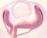
For embryological formation of the retina, click on the thumbnail images at left.
Comments and questions: dgking@siu.edu
SIUC / School
of Medicine / Anatomy / David
King
https://histology.siu.edu/ssb/EE020b.htm
Last updated: 29 July 2023 / dgk