

Comparison of peripheral and central retina
 |
 |
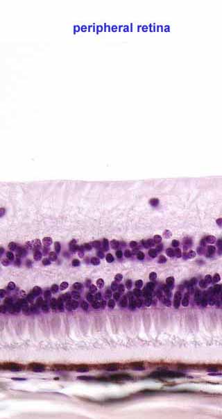
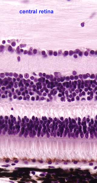
SSB IMAGE INDEXThese two sections, through regions of the retina that are peripheral (farther from the macula) and central (closer to the macula), are aligned at the pigmented epithelium (cuboidal cells with brownish color, near bottom).
Note that "inner" and "outer" in the names of retinal layers refer to the eyeball, not to the surface of the retina. Thus "inner" is upward in these two images, while "outer" is downward.Note that the peripheral retina is not only much thinner than the central retina but also has many fewer cells (as indicated by the number of nuclei). This is most apparent in the ganglion cell layer (the topmost layer of nuclei, containing relatively few ganglion cells) and in the inner nuclear layer (the middle layer of nuclei, containing bipolar cells). Density of receptor cells also varies regionally but less markedly so than for the integrative bipolar and ganglion cells.
The resolution of the retina (i.e., its ability to distinguish fine details of the image) is limited not so much by the absolute number of receptor cells as by the number of ganglion cell axons which pass image "pixels" along the optic nerve. Thus the resolution is much higher in the central retina, particularly in or near the macula, where the number of ganglion cells is much greater than in the peripheral retina.
Comments and questions: dgking@siu.edu
SIUC / School
of Medicine / Anatomy / David
King
https://histology.siu.edu/ssb/NM012b.htm
Last updated: 29 July 2023 / dgk