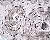
Each osteon consists of parallel, concentric lamellae surrounding a Haversian canal.
Highlighted in blue are the cement lines which mark the boundaries of present and former osteons.
These lines indicate the shape of the surface upon which osteoblasts began laying down fresh lamellae after osteoclasts had excavated a channel throught the bone.
The patterns of lamellae reflect the history of bone remodelling. Osteons with complete concentric lamellae are the most recently formed. Other lamellae, which terminate abruptly against a cement line, are the fragmentary remains of former osteons that have been partially removed and replaced during remodelling.
- Number 1 indicates a recently formed osteon.
- Number 2 indicates a somewhat older osteon, as demonstrated by the encroachment of osteon 1.
- Number 3 indicates interstitial lamellae, representing the remnants of still older osteons.
Click on a thumbnail below for additional information.
Comments and questions: dgking@siu.edu
SIUC / School
of Medicine / Anatomy / David
King
https://histology.siu.edu/ssb/NM035b1.htm
Last updated: 13 August 2021 / dgk