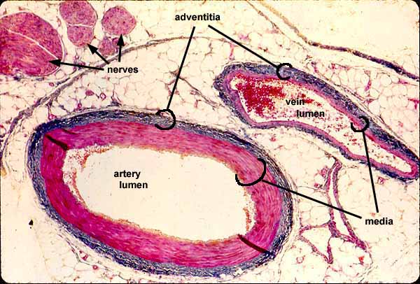

Artery, vein, nerve trichrome stain

CARDIOVASCULAR IMAGE INDEXPeripheral arteries, veins, and nerves tend to travel and branch in parallel. Wherever one of these structures is found, the other two are likely to be close by.
In this trichrome stained specimen, collagen is colored blue and smooth muscle is red. Red blood cells in the venous lumen are brighter red. The background is adipose connective tissue.
Note differences between artery and vein, not only the thickness of the vessel wall relative to the lumen but also the overall shape (artery rounder, vein flatter).
- The intima is not noticable at this magnification.


- The media is the thickest, most conspicuous layer of the artery, much less pronounced in the vein.
- In this loose connective tissue, the adventitia comprises distinct layer.
Comments and questions: dgking@siu.edu
SIUC / School
of Medicine / Anatomy / David
King
https://histology.siu.edu/crr/CR020b.htm
Last updated: 22 May 2022 / dgk