RENAL IMAGE INDEX
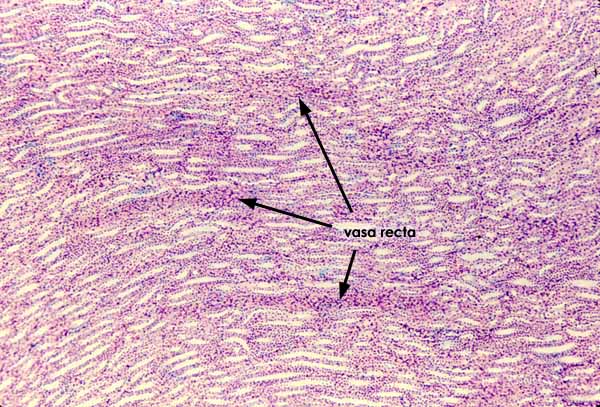
This image illustrates the mid-region of the renal medulla. Note that in contrast to cortex (thumbnail at right), the medulla contains no renal corpuscles and all tubules are approximately parallel to one another.
Vasa recta appear as tightly packed bundles of vessels.
In this specimen, most tubules are cut lengthwise, indicating that the plane of section is perpendicular to the cortex. To view a similar region in a different plane of section, click on the thumbnail at right..
To view medullary tubules at higher magnification, click on one of the thumbnails below.
Comments and questions: dgking@siu.edu
SIUC / School
of Medicine / Anatomy / David
King
https://histology.siu.edu/crr/RN014b.htm
Last updated: 30 May 2022 / dgk