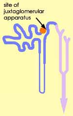
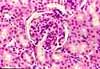
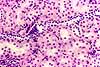
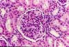
Juxtaglomerular cells, renal cortex
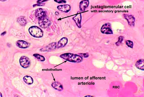
This image from a thin (1µm) section shows sufficient detail to resolve secretory granules within juxtaglomerular cells, found in the wall of an afferent arteriole. (The clear white "bubbles" near the vascular endothelium and at upper right are preparation artefacts.)
For additional views of the juxtaglomerular apparatus, click on one of the thumbnails below.
RENAL IMAGE INDEX / RENAL STUDY GUIDE
Comments and questions: dgking@siu.edu
SIUC / School
of Medicine / Anatomy / David
King
https://histology.siu.edu/crr/RN108b.htm
Last updated: 16 September 2021 / dgk