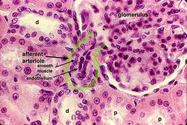
This image of renal cortex includes several sections of proximal (p) and distal (d) tubules and the region around the vascular pole of one renal corpuscle.
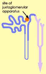
The distal tubule near the afferent arteriole (outlined in green) is presumably approaching the juxtaglomerular apparatus, although the macula densa is not visible in this plane of section.
For lower magnification, click on the thumbnail at left.
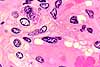
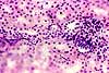
For additional views of the juxtaglomerular apparatus, click on one of the thumbnails below.
RENAL IMAGE INDEX / RENAL STUDY GUIDE
Comments and questions: dgking@siu.edu
SIUC / School
of Medicine / Anatomy / David
King
https://histology.siu.edu/crr/RN037b.htm
Last updated: 14 September 2021 / dgk