
Notes
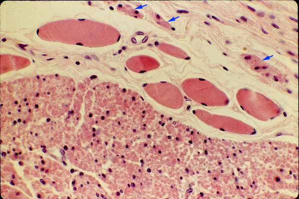
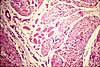
The muscularis externa consists of skeletal muscle in the proximal (upper) third of the esophagus and smooth muscle in the distal third. This micrograph shows a transitional region in which both types of muscle are present.
In this figure, a small amount of connective tissue occupies the spaces between muscle fibers. In addition to the large mass of smooth muscle in the lower half of the figure, some inconspicuous small bundles of smooth muscle may be seen near the top center and the upper right (indicated by blue arrows).
The appearance, or visual texture, of muscle varies dramatically depending on the orientation of the fibers with respect to the plane of section. All of the muscle fibers in this view are cut in cross section.
Individual smooth muscle fibers are usually difficult to resolve, even at fairly high magnification. Cross-sectioned smooth muscle displays only scattered nuclei. In each individual fiber, the nucleus occupies only a small fraction of the fiber's length. So nuclei appear only in a similarly small proportion of the fibers cut by any single section. (Remember, each smooth muscle is a single cell, each with its own nucleus.)
Individual striated muscle fibers are conspicuous as individual units. Each fiber has so many nuclei that any random cross section typically displays at least one and often several, normally located around the periphery of the fiber.
Related examples:
 |
 |
 |
 |
 |
|
Comments and questions: dgking@siu.edu
SIUC / School
of Medicine / Anatomy / David
King
https://histology.siu.edu/erg/GI004b.htm
Last updated: 9 May 2022 / dgk