

Notes
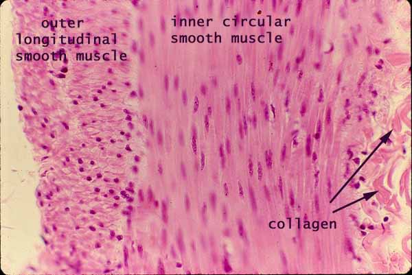
The muscularis externa of the intestine consists of smooth muscle fibers arranged into an inner circular layer and an outer longitudinal layer.
The appearance of smooth muscle varies dramatically, depending on the orientation of the fibers with respect to the plane of section.
In this section, the circular muscle fibers are cut longitudinally, while the longitudinal muscle fibers are cut in cross section (transversely).
Note especially the appearance of smooth muscle nuclei. These nuclei appear small and round when cut in cross section, very long (and often wiggly) in longitudinal section, and somewhere in between in oblique section.
In cross-sectioned smooth muscle, only a fraction of the fibers display nuclei. Recall that each smooth muscle fiber is a single cell, each with its own nucleus. In each such cell, the nucleus occupies only a small proportion of the cell's length. So nuclei appear only in a similarly small proportion of the fibers cut by any single section.
Collagen resembles smooth muscle, in being fibrous and eosinophilic (i.e., pink staining with H&E). However, note in the thumbnail to right how distinctly different connective tissue appears, due to the collagen fibers' irregular size and orientation, and by the haphazard shape and distribution of fibroblast nuclei.
Smooth muscle fibers usually occur in bundles in which all fibers have approximately the same size and orientation.
Collagen fibers, in contrast, tend to be various in size and orientation (except in specialized cases, such as tendons).
Subtle differences in color can sometimes distinguish smooth muscle from collagen in specimens stained with H&E, but the quality of difference varies from specimen to specimen. Texture (size, shape, orientation of fibers and associated cell nuclei) is much more reliable. When it is important to make this distinction quickly and reliably, a stain that is selective for collagen may be chosen (e.g., one of the trichrome stains).
Nervous tissue of Auerbach's plexus is not apparent on this micrograph.
Related examples:
 |
 |
 |
 |
 |
|
Comments and questions: dgking@siu.edu
SIUC / School
of Medicine / Anatomy / David
King
https://histology.siu.edu/erg/GI012b.htm
Last updated: 14 May 2022 / dgk