
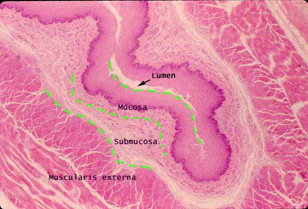
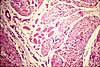
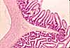
Esophagus, basic layers (cross section)
Notes
The basic layers of the GI tract are especially distinct in the esophagus.
In the esophageal mucosa --
Esophageal epithelium is non-keratinized stratified squamous.
Esophageal lamina propria is less cellular (fewer lymphocytes) than that in the stomach and intestine, presumably because the protective stratified squamous epithelium is more effective at keeping out foreign antigens.
Esophageal muscularis mucosae is noticably thicker than that in the stomach and intestine, and includes only longitudinal muscle fibers. At this magnification, the muscularis mucosae is not conspicuous.
Connective tissue of the submucosa is typically more fibrous and less cellular than that of the lamina propria of the mucosa. Esophageal submucosa includes scattered esophageal glands (not included in this micrograph, see this image) and a venous plexus (not conspicuous in this micrograph).
Muscularis externa of the esophagus consists of the standard inner circular and outer longitudinal layers of smooth muscle, with Auerbach's plexus in between. Both layers are included here.
Note that muscle fibers appear to be cut in cross section in the inner layer and in longitudinal section in the outer layer. This indicates that the esophagus itself must have been cut longitudinally (parallel to the axis of the tube). Then the specimen must have been folded to give the shape seen above.
The vagina provides an instructive contrast with the esophagus. The vagina and the esophagus are both tubular organs with a mucosa lined by non-keratinized stratified squamous epithelium. Hence these two appear superficially similar. However, bundles of smooth muscle are interwoven with connective tissue throughout the wall of the vagina, so that there is no clear demarcation of submucosal and muscularis layers.
Certain special features which distinguish these organs (e.g., glycogen storage in vaginal epithelium, submucosal glands in the esophagus) are not always apparent in routine sections. So the difference between "layers distinct" and "layers not distinct" may be the easiest and most reliable way to distinguish these two organs (e.g., on a practical evaluation).
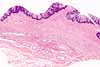
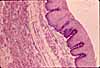
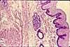
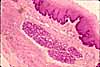
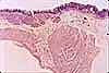
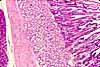
| More esophagus examples: | ||||||
 |
 |
 |
 |
|||
| Basic
layers in other regions of the GI tract |
||||||
 |
 |
 |
Comments and questions: dgking@siu.edu
SIUC / School
of Medicine / Anatomy / David
King
https://histology.siu.edu/erg/GI014b.htm
Last updated: 27 May 2022 / dgk