
Prostate
INTRODUCTION
Please note: Any student who calls this the "prostrate" gland will have her/his rubber glove confiscated.
The prostate is a small gland designed to impose humility on aging men. By virtue(?) of its remarkable location, the prostate assumes an unfortunate clinical significance out of all proportion to its size and normal function. In case you haven't already learned, BPH (Benign Prostatic Hyperplasia, or enlargement of the prostate; example from WebPath) is a standard concomitant of advancing age in men. Since the male urethra passes through the prostate, and since the organ has a fibrous capsule, BPH typically compresses the urethra and thereby interferes with urination.
Furthermore, the prostate is especially prone to malignant neoplasm. Prostate cancer is one of the leading causes of cancer death among men. Histologic diagnosis is typically made on the basis of scrapings (via the the urethra) or needle biopsy (through the rectum), both of which require some considerable familiarity with the normally various appearance of prostatic tissue.
You are encouraged to visit the WebPath tutorial on the prostate, for an accessible, illustrated introduction to prostate pathology. This site sholud be better appreciated after you are acquainted with the appearance of the normal prostate.
"The biologic role of the prostate calls for the slow accumulation and occasional rapid expulsion of small volumes of fluid. These requirements are optimally met by a muscular organ having a large storage capacity and low secretory capacity" (J.E. McNeal, in Sternberg, Histology for Pathologists, 2nd edition, Lippencott-Raven, 1997).
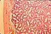
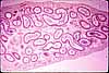
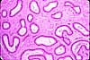
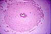
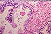
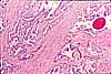
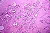
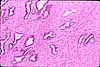
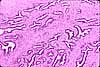
The appearance of prostatic glandular tissue varies considerably from site to site within the organ (there are several different zones), but all appear rather irregularly tubular with a convoluted epithelium lined by columnar, secretory cells. The secretory tissue is embedded in an extensive fibromuscular stroma that contains both collagen and smooth muscle, thoroughly interwoven. Glandular lumens may normally contain small acidophilic concretions called corpora amylacea.
Multiple images are included here to provide a sense of diversity. The same basic features appear in each image, but the varied appearance of several images should be examined.
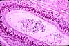
The urethra is, of course, the tube which extends from the bladder through (in men) the prostate and thence out through the penis. As you should remember from CRR, both the bladder and the urethra have a muscular wall and a mucosal lining with a transitional epithelium (urothelium), a stratified epithelium in which both cell shape and number of layers can change markedly during the normal process of distension. (The urethra shown in the thumbnail image appears to be a female urethra; it is surrounded neither by prostate nor by penile erectile tissue.)
Compression of the urethra by BPH (Benign Prostatic Hyperplasia, or enlargement of the prostate) can cause urinary difficulties.
 |
 |
 |
 |
 |
 |
 |
 |
 |
 |
 |
 |
|
 |
 |
 |
 |
 |
 |
 |
 |
 |
|
 |
SIUC / School
of Medicine / Anatomy / David
King
https://histology.siu.edu/erg/prostate.htm
Last updated: 2 February 2023 / dgk