
Notes
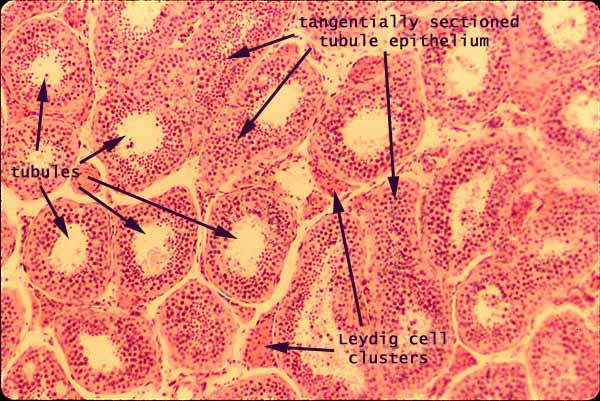
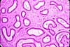
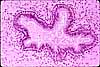
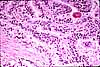
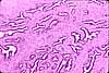
The testis consists primarily of seminiferous tubules.
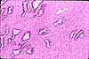
The complex appearance of the tubule epithelium results from the presence, in addition to columnar epithelial Sertoli cells, of germ cells undergoing meiosis.
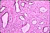
The organization of tubule epithelium appears most clearly when individual tubules are cut in cross section. Tangential sections through tubule epithelium (as indicated in the micrograph above) present a solid appearance with cell structure varying from place to place depending on the depth at which the epithelium was cut.
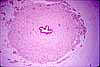
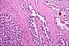
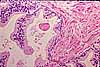
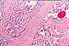
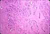
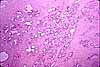
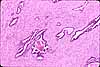
Between the tubules is a delicate connective tissue stroma, or interstitial tissue, containing clusters of testosterone-producing Leydig cells.
Leydig cells may be recognized not only by their location within the testicular interstitium but also by their round nuclei and extensive acidophilic cytoplasm.
Related examples:
 |
 |
 |
|
 |
 |
 |
 |
 |
 |
 |
 |
 |
 |
 |
 |
 |
 |
 |
 |
 |
 |
 |
 |
Comments and questions: dgking@siu.edu
SIUC / School
of Medicine / Anatomy / David
King
https://histology.siu.edu/erg/RE026b.htm
Last updated: 18 May 2022 / dgk