Notes
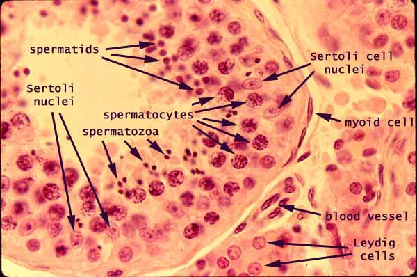
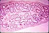
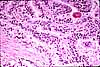
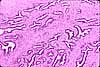
Within the seminiferous tubules of the testis, the complex appearance of the tubule epithelium results from the presence of germ cells undergoing meiosis nestled among the columnar epithelial Sertoli cells.
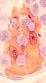
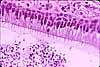
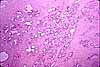
Sertoli cells
Sertoli cells have a complex shape (image at right), enveloping germ cells in various stages of meiosis and sperm maturation. Sertoli cell nuclei may be recognized by their typical epithelial shape and texture -- oval, euchromatic, usually with a prominent nucleolus -- in contrast with the round nuclei and condensed chromatin of the various germ cells.
Any particular section of tubule epithelium represents a "snap-shot" of germ cells at one arbitrary moment during the process of sperm cell formation.
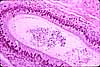
- Large round nuclei nearer the base of the epithelium belong to cells early in the process of meiotic cell division -- spermatogonia, primary spermatocytes. Nuclear texture in these cells depends on the precise stage of meiosis.
- Nuclei nearer the lumen belong to concluding stages of meiosis -- secondary spermatocytes, spermatids, and spermatozoa.
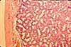
Various segments around the circumference of a tubule display different phases in the repeating cycle of meiosis, with all of the cells within one segment being "in phase."
In the micrograph above, note how the germ cells nearest the the lumen vary from segment to segment. As indicated at right, these cells are secondary spermatocytes in segment 1, spermatids in segment 2, and spermatozoa in segment 3.
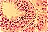
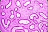
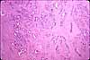
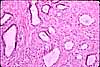
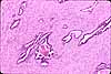
Between the tubules is a delicate connective tissue stroma, or interstitial tissue, containing clusters of testosterone-producing Leydig cells.
Leydig cells may be recognized not only by their location within the testicular interstitium but also by their round nuclei and extensive acidophilic cytoplasm.
Contractile myoid cells surround each tubule.
Related examples:
 |
 |
 |
 |
 |
 |
 |
 |
 |
 |
 |
 |
 |
 |
 |
 |
 |
 |
 |
 |
 |
 |
 |
 |
Comments and questions: dgking@siu.edu
SIUC / School
of Medicine / Anatomy / David
King
https://histology.siu.edu/erg/RE028b.htm
Last updated: 2 February 2023 / dgk