
Skin, hypodermis

Notes
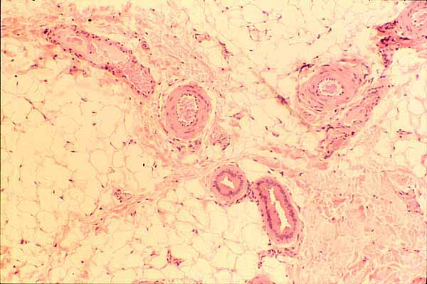
This image shows connective tissue deep in skin. Click on the image to identify individual features.
Subcutaneous adipose tissue occupies much of the image. Fibrous connective tissue appears at lower right.
Conspicuous features include several blood vessels, and a portion of a sweat gland.

The pink color in this image represents eosinophilic material, in this case mostly one of the following:
- Most of the pink material represents extracellular collagen fibers.
- The somewhat deeper pink encircling the arteries is the cytoplasm of smooth muscle cells (thumbnail at near right).
- The deeper pink of the sweat gland is cytoplasm of sweat gland epithelial cells (thumbnail at far right).
The pale background color represents unstained material, in this case one of the following:.
- Larger, irregular open spaces represent the lipid droplets within adipocytes. (Intact adipocytes appear round, with continuous margins. Adipocytes which have been damaged during specimen preparation appear as irregular shapes with broken margins.)
- Within the sweat gland, the clear space represents the glandular lumen, continuous with the outside world.
- Elsewhere, the thin interconnected pale spaces between collagen fibers represent connective tissue ground substance.
The very dark purple (nearly black) spots are cell nuclei. At this resolution, they cannot be individually identified except by context.
- Nuclei adjacent to the lumen of a blood vessel belong to vascular endothelial cells.
- Nuclei in the muscular wall of an artery may belong to smooth muscle cells.
- Irregular nuclei scattered at random among collagen fibers belong mostly to fibroblasts Some may also belong to macrophages, mast cells, or capillary endothelial cells.
- Occasionally a thin nucleus adjacent to a large lipid droplet might belong to an adipocyte. More probably, such nuclei might belong to fibroblasts or to capillary endothelium located between the adipocytes.
Comments and questions: dgking@siu.edu
SIUC / School
of Medicine / Anatomy / David
King
https://histology.siu.edu/intro/IN027b.htm
Last updated: 13 June 2022 / dgk