
Hair Follicles in Skin

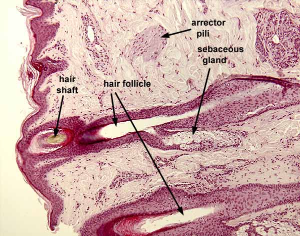
This image shows hair follicles near the surface of skin, surrounded by fibrous connective tissue of the dermis.
A hair follicle is a tubular invagination lined by stratified squamous epithelium similar to epidermis with a central lumen that might (or might not) contain a hair shaft. Typically each hair follicle is associated with a sebaceous gland (not always visible in the same plane of section) which opens into the lumen.
The hair shaft is attached only in the bulb at the bottom of the follicle, so when the tissue is sliced to prepare a slide, the sectioned bit of hair may fall out.
The appearance of hair follicles varies considerably, not only with plane of section but also with depth in the dermis and also with growth phase. Consult a histology text for details.
Each hair follicle is associated with a small bundle of smooth muscle, the arrector pili, which originates in the upper dermis and inserts on the connective tissue surrounding the base of the follicle. Contraction of this muscle causes the hair to stand erect.
Much of the area of this this image is occupied by connective tissue of the dermis, consisting of collagen (pink), ground substance (pale background), and scattered fibroblasts and other connective tissue cells (dark).
Deeper in the skin at the base of each hair follicle (thumbnail at right), is the hair bulb where the hair shaft is attached and where the hair shaft is produced by proliferation of epithelial cells of the bulb.
Comments and questions: dgking@siu.edu
SIUC / School
of Medicine / Anatomy / David
King
https://histology.siu.edu/intro/IN036b.htm
Last updated: 12 June 2022 / dgk