Click on one of the thumbnails at right for low-magnification overview.
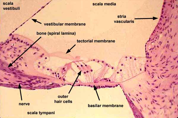
Hair cells of the organ of Corti comprise two groups, the outer hair cells which are conspicuous on this image and the inner hair cells which are less conspicuous here, located above the tip of the bony spiral lamina.
Historical note: The entire structure of the organ of Corti, spanning this image from left to right, includes several unlabelled structural details, named eponymously for Deiters, Claudius, Hensen, Boettcher, and Nuel. These are visible here as many of the various unlabelled cells on either side of the labelled outer hair cells.Hair cells of the organ of Corti are stimulated by relative movement of the tectorial membrane and the basilar membrane, driven by pressure waves between scala vestibuli and scala tympani.
The space of the scala media, including the spaces surrounding the hair cells, are filled with endolymph, secreted by cells of the stria vascularis. (Blood vessels between the cuboidal cells of the stria vascularis are not readily noticable in this image.)
The vestibular membrane, separating the scala vestibuli from the scala media, is also called Reissner's membrane (commemorating Ernst Reissner, b. 1824).Scala vestibuli and scala tympani are filled with perilymph.
Axons of the nerve within the spiral lamina pass by the spiral ganglion (where their cell bodies reside), hence into the modiolus and then the auditory nerve.
Comments and questions: dgking@siu.edu
SIUC / School
of Medicine / Anatomy / David
King
https://histology.siu.edu/ssb/EE001b.htm
Last updated: 5 July 2022 / dgk