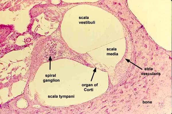
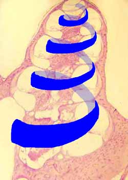
A section across one turn of the spiral cochlea reveals structures which extend lengthwise through all the turns of the cochlea, from its base at the modiolus to its apex at the helicotrema, as indicated by the blue helix in the overview. (NOTE: This is NOT a human cochlea, which is shorter and broader, with fewer turns.)
Click on one of the thumbnails at right for higher- or lower-magnification views.
The space of the scala media, including the spaces surrounding the hair cells, are filled with endolymph.
- Endolymph is secreted by cells of the stria vascularis.
Scala vestibuli and scala tympani are filled with perilymph.
Sensory axons stimulated by hair cells of the organ of Corti pass by the spiral ganglion (where their cell bodies reside), hence into the modiolus (the central column of the cochlea) and the auditory nerve.


