Ear
 Regions of the ear.
REGIONS of the ear
The ear has three distinct regions -- outer ear, middle ear, and inner ear.
- The outer ear includes the pinna (the visible ear, consisting mostly of skin and cartilage) and the ear canal. The latter is lined by keratinized stratified squamous epithelium. This lining differs from skin by the presence of specialized ceruminous (ear-wax) glands.
- The middle ear is basically a space, communicating via the eustacian tube with the oropharynx. It is lined by a very thin non-keratinized stratified squamous epithelium. Spanning the space of the middle ear are the three middle ear bones, the malleus (hammer), incus (anvil), and stapes (stirrup).
- The eardrum is a thin membrane separating the outer ear from the middle ear. It is sandwich of tissues, with keratinized stratified squamous epithelium facing the outer ear, non-keratinized stratified squamous epithelium facing the middle ear, and a very thin layer of connective tissue in between.
- The inner ear is the portion of the ear which contains sensory receptors. The remainder of this study guide describes the inner ear.
OVERVIEW of the INNER EAR
The inner ear has a complex structure. The following basic concepts should help organize that complexity.
The inner ear is located within the bony labyrinth (more below). It contains complex sensory receptors serving both balance and hearing.
Image courtesy Alec Salt, used with permission.
- Linear acceleration (including gravity, for head orientation) is sensed by the otolith organs of the saccule and the utricle.
- Angular acceleration (i.e., head rotation) is sensed by the cristae ampularis of the semicircular canals.
- Hearing is sensed by the organ of Corti within the scala media of the cochlea.
- All of these several senses of the inner ear utilize the same mechanoreceptor cell type, epithelial hair cells.
- Hair cells are housed within an elaborately-shaped chamber called the membranous labyrinth.
- The membranous labyrinth is filled with a unique fluid called endolymph, secreted by cells of the stria vascularis. Endolymph differs substantially from all other fluids of the body and provides a special fluid environment for the hair cells
- The membranous labyrinth provides interconnection among the cochlea, saccule, utricle, and semicircular canals.
- The membranous labyrinth is housed within the bony labyrinth.
- The space within the bony labyrinth which surrounds the membranous labyrinth is filled with a fluid called perilymph.
- The distinction between endolymph and perilymph is physiologically significant; see below.
BONY LABYRINTH and MEMBRANOUS LABYRINTH
Image courtesy Alec Salt, used with permission.
The inner ear resides within a space called the bony labyrinth.
- The oval window forms a potential opening from the middle ear into the bony labyrinth.
- The stapes of the middle ear plugs this opening; but . . .
- The stapes is flexibly attached and can vibrate to transmit pressure waves to the fluid that fills the bony labyrinth.
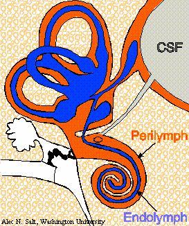
(Sound is carried from the eardrum across the middle ear by the three middle ear ossicles, ending with the stapes at the oval window.)Suspended within the bony labyrinth, and approximating its shape, is an interconnected set of membrane-lined chambers and passageways called the membranous labyrinth.
- In the diagram at right, the color orange occupies the space of the bony labyrinth, while the membranous labyrinth is blue.
- The name labyrinth suggests the complex shape of these chambers and passageways.
- Three semicircular canals comprise looping tubules which leave and return to the vestibule.
- Within each semicircular canal of the bony labyrinth is a semicircular canal of the membranous labyrinth.
- The saccule, the utricle, and the semicircular canals together comprise the vestibular system.
- The cochlea is shaped like a snail-shell which spirals away from the vestibule.
- A single coiled tunnel of the bony labyrinth is subdivided into three levels ("scalae") by membranes of the membranous labyrinth.
- The portion of the membranous labyrinth within the cochlea is called the scala media.
- The scala media of the cochlea is connected to the saccule by the ductus reuniens, or Hensen's duct [after Victor Hensen, b. 1835].
The membranous labyrinth is filled with a unique fluid called endolymph (blue in diagram). Surrounding the membranous labyrinth (i.e., filling the remaining space of the bony labyrinth) is a fluid called perilymph (orange in diagram).
HAIR CELLS
Hair cells are the specialized mechanoreceptor cells of the auditory and vestibular systems. Hair cells are found in several positions along the chambers and passageways of the membranous labyrinth.
- Hair cells are basically columnar epithelial cells.
- At the apical end of each hair cell is a set of "hairs" (cytoplasmic projections, kinocilium and stereocilia) embedded in a mass of extracellular jelly.
- At the basal end of each hair cell are synapses onto sensory axons.
- Hair cells work similarly throughout the inner ear.
- A hair cell responds when movement of the extracellular jelly displaces its kinocilium and stereocilia. Displacement is excitatory in one direction (toward the kinocilium) and inhibitory in the opposite direction.
- Displacement of the kinocilium and stereocilia alters conductance of ion channels, in turn affecting release of neurotransmitter onto the associated sensory axon. (These axons project along the auditory and vestibular nerves, cranial nerve VIII).
- Hair cells function within a fluid environment, the endolymph, with a unique ionic composition.
- The process of sensory transduction in hair cells (i.e., the conversion of an external stimulus, in this case small movement, into neural activity) has been intensively investigated. For detailed information, consult your print resources (e.g., Kandel et al.)
- The mechanical disposition of the jelly in relation to the spaces of the membranous labyrinth determines how hair cells respond.
- Hair cells in the semicircular canals respond to angular acceleration (rotation).
- Hair cells in the in the otolith organs respond to linear acceleration.
- Hair cells in the organ of Corti of the cochlea respond to sound.
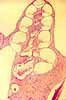
OTOLITH ORGANS (SACCULE and UTRICLE)
The saccule and utricle contain patches of hair cells called maculae ("macula" means "spot" or "patch").
- A small mass of jelly rests on top of the hair cells of the macula.
- In this jelly are numerous tiny mineral concretions, called otoliths ("earstones" or "earsand").
- Clinical Note: If any otoliths break loose (e.g., due to head trauma), they may come to rest in an inappropriate place, stimulating the hair cells in a semicircular canal) and cause disturbance in balance (e.g., see benign paroxysmal positional vertigo, or BPPV).
- Hair cells of the macula are deflected by the weight or inertia of the otoliths. Together the two pairs of otolith organs (one of each in each ear) can sense head orientation (gravity) or linear acceleration in any direction.
Image courtesy Alec Salt, used with permission.
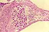
SEMICIRCULAR CANALS
Each semicircular canal of the bony labyrinth is a hollow passageway looping out from and back to the vestibule. Within each of these passageways is a semicircular canal of the membranous labyrinth.
- At one end of each membranous semicircular canal is a small enlargement called the ampulla.
Within each ampulla is a ridge or "crest" called the crista.
- The crista is covered with hair cells.
- A small mass of jelly, called the cupola ("cap") rests on top of the hair cells of the crista.
Hair cells of the ampullae respond to angular acceleration (i.e., rotation of the head).
- There are three semicircular canals in each ear, oriented in three mutually-perpendicular planes.
- Rotation of the head in any direction will cause inertial fluid movement in one or more of the semicircular canals.
- Fluid motion in a semicircular canal pushes the the cupola like a swinging door.
- Movement of the cupola in turn deflects the projections of the hair cells.
Clinical Note: Should loose otoliths enter a semicircular canal, they may stimulate the hair cells inappropriately and cause disturbance in balance (see benign paroxysmal positional vertigo, or BPPV).The planes of orientation of the semicircircular canals correspond to the planes of action of the extraocular muscles, allowing simple reflexes to coordinate eye movement with head rotation. (Amazingly, at least to this writer, this correspondence has been maintained through evolution even while position and orientation of both eyes and ears have been modified.)
Image courtesy Alec Salt, used with permission.
COCHLEA
The cochlea houses an elaborate configuration of membranous labyrinth and hair cells, called the organ of Corti, designed for auditory reception.
The basic shape of the cochlea is that of a snail-shell, or tapering helix.
NOTE: The human cochlea is short and broad, with fewer turns than shown here. Micrographs at this website (as well as in many other references) show the cochlea of a laboratory rodent which is proportionately taller and narrower.
The spiraling tunnel (blue, in image at right) that forms the cochlea of the bony labyrinth is divided into three distinct channels by portions of the membranous labyrinth attached to bony ridges. Each of these channels is called a "scala," meaning "ramp" or "incline" (think of a musical "scale").
- The scala vestibuli ascends from the vestibule (hence vestibuli in the name) to the tip of the cochlea.
- The scala vestibuli contains perilymph.
- At the tip of the cochlea, the scala vestibula connects to the scala tympani at the helicotrema.
- The scala tympani descends from the tip of the cochlea to the round window. (There is an elastic energy-dissipating membrane covering the round window (hence tympani in the name).
- The scala tympani, like the scala vestibuli, contains perilymph.
- At the tip of the cochlea, the scala vestibula and the scala tympani are connected through the helicotrema.
The scala media, also called the cochlear duct, lies along the length of cochlear spiral, in a "middle" position between the scala vestibuli and scala tympani.
Image courtesy Alec Salt, used with permission.
- The scala media contains endolymph, secreted by the stria vascularis. The stria vascularis resembles a stratified cuboidal epithelium lining the outer curve of the scala media. But unlike any proper epithelium (and as the name vascularis suggests) this tissue contains capillaries among the cuboidal cells.
- The organ of Corti lies within the scala media.
- The scala media is separated from the scala vestibuli by the very thin vestibular membrane, also called Reissner's membrane (commemorating Ernst Reissner, b. 1824).
- The scala media is separated from the scala tympani by the basilar membrane.
Clinical note: A cochlear implant is inserted into the scala tympani, where it lies close to the organ of Corti and can artificially stimulate axons of the auditory nerve.
The central column (the modiolus) of the helical cochlea contains axons serving the organ of Corti on their way to the auditory nerve. A bony ridge, the spiral lamina, extends out from the modiolus and provides support for the organ of Corti. A tubular cavity within the spiral lamina (Rosenthal's canal, named after F.-C. Rosenthal, b. 1780) contains the cell bodies of the axons of the auditory nerve. Because this collection of nerve cell bodies has a helical shape paralleling the cochlear scalae, it is called the spiral ganglion.
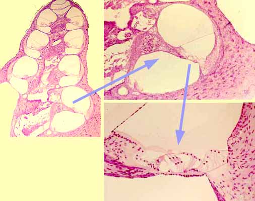
ORGAN OF CORTI
The organ of Corti (commemorating Alfonso Corti, b. 1822) is an elaborate structure with more named parts than the rest of inner ear altogether. Several key features are listed below. Click on an image for an enlarged view.
The organ of Corti is contained within the scala media.
- The organ of Corti is a long strip of tissue that extends the length of the scala media, from the base of the cochlea to its apex.
- The organ of Corti is usually illustrated in cross-section, as it is on this page. In contrast, tissue sections on microscope slides typically contain several differing appearances of the organ of Corti, as the organ is sliced somewhat differently in each turn of the helix.
- The fluid environment for the organ of Corti is endolymph, which fills the scala media. (Endolymph is secreted by cells of the stria vascularis.)
- Within the complex strip of tissue that comprises the organ of Corti are specialized sensory hair cells.
- The entire complex (the whole organ of Corti) rests on the basilar membrane of the cochlea.
- This basilar membrane supports the basal ends of the hair cells in the organ of Corti.
- The apical ends of hair cells touch the tectorial membrane, a "shelf" of jelly that is supported immovably on the spiral lamina.
When the basilar membrane flexes in response to sound waves (i.e., to pressure waves delivered to inner-ear fluid by the middle-ear ossicles), the organ of Corti, including its hair cells, also moves.
- Thus, when the basilar membrane is moved by pressure waves (i.e., by sound), the hair cells move relative to the tectorial membrane, causing stimulatory deflection of the apical ends of the hair cells.
Clinical note: A cochlear implant is inserted into the scala tympani, where it lies close to the organ of Corti and can artificially stimulate axons of the auditory nerve.
The organ of Corti is considerably more complex than this simple account implies, with, among other things, two functionally distinct classes of hair cells (inner and outer). Synapses from the inner hair cells apparently supply most of the sensory information that goes to the brain, while the outer hair cells (the ones which are most readily recognized by light microscopy) have a curious mechanical function. (For more, see for example Inner and Outer Hair Cells, Baylor College of Medicine.)
Historical note: Among the many histological details associated with the organ of Corti are eponymous structures commemorating Rosenthal, Reissner, Deiters, Claudius, Hensen, Boettcher, and Nuel.Kölliker's organ, the embryonic precursor of the hair cells in the organ of Corti, may be the only eponymous structure in human anatomy to commemorate Albert von Kölliker, "the father of modern histology," who first described it in 1863.
Image courtesy Alec Salt, used with permission. ENDOLYMPH and PERILYMPH
The membranous labyrinth is filled with endolymph and surrounded by perilymph.
The distinction between endolymph and perilymph is physiological significant.
Endolymph (blue, in image at right) is a unique fluid, with high K+ concentration and very low Na+ concentration. This endolymph provides the appropriate ionic environment for hair cell function.
Endolymph is secreted by cells of the stria vascularis, along the scala media of the cochlea. The stria vascularis resembles a stratified cuboidal epithelium, but unlike any proper epithelium (and as the name vascularis suggests) this tissue contains capillaries among the cuboidal cells.
Endolymph is produced continuously. The outlet (or "drain") for removing endolymph is via the vestibular aquaduct into the endolymphatic sac, wherefrom endolymph moves into cerebrospinal fluid.
This is one of three sites associated with the nervous system where a special fluid is produced by a unique tissue, with this fluid needing an outlet elsewhere to avoid buildup of pressure. (The other two sites are the eye and the brain. In the eye, aqueous humor secreted by ciliary processes is drained through the canal of Schlemm. In the brain, cerebrospinal fluid secreted by choroid plexus is drained through arachnoid villi into the superior sagittal sinus.) In each of these sites, an imbalance between production and drainage can cause neurological symptoms.
Perilymph (orange, in image at right) is similar to ordinary interstitial fluid. Perilymph fills the spaces of the bony labyrinth surrounding the membranous labyrinth.
In the vestibular system (surrounding the saccule, utricle, and semicircular canals), perilymph simply provides a cushioning support for the membranous labyrinth.
In the cochlea, perilymph of the ascending scala vestibuli and the descending scala tympani conveys pressure waves (sound) across the scala media. Pressure waves flex the basilar membrane and thereby stimulate hair cells of the organ of Corti.
Comments and questions: dgking@siu.edu
SIUC / School
of Medicine / Anatomy / David
King
https://histology.siu.edu/ssb/ear.htm
Last updated: 22 July 2023 / dgk