Like the eyeball itself, the eyelid displays considerable histological complexity.
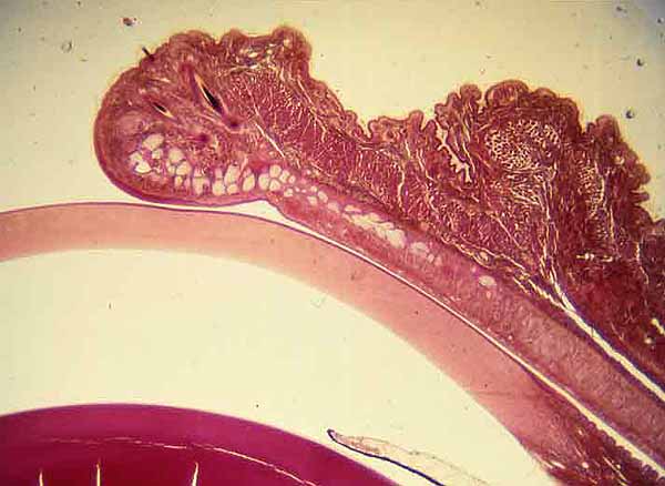
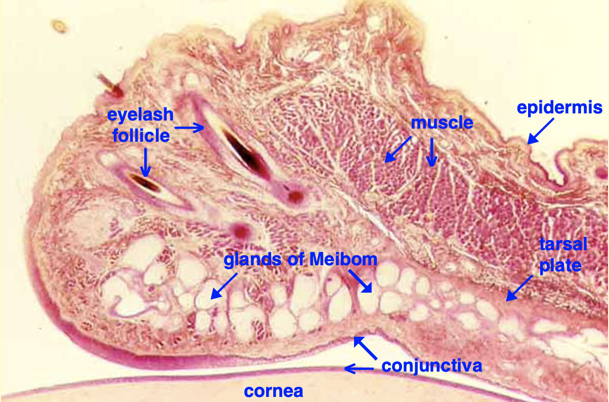
This section displays the very thin epidermis of the eyelid, eyelash hair follicles, skeletal muscle, and sebaceous Meibomian glands within the tarsal plate, as well as conjunctival epithelium of both eyelid and cornea. (The Meibomian glands are over-exposed in this image, obscuring their sebaceous appearance.)

For lower magnification, click on thumbnail at right.
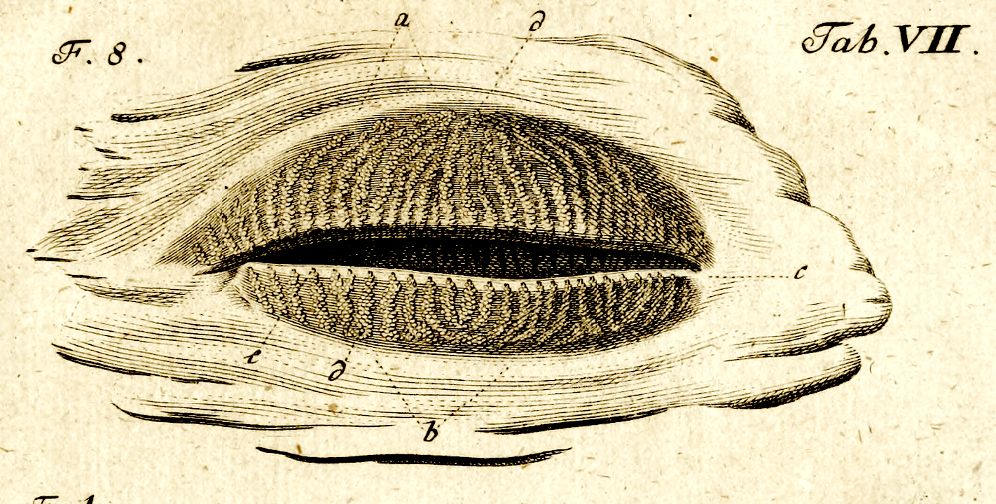
Below is an image of Meibomian glands on the inside of the eyelid, from a 1780 edition of Johann Zinn's atlas of eye anatomy (accessed at the Wellcome Collection). For an even earlier view of Meibom's glands, see Meibom, 1666.


