
William Bowman's eye anatomy (1847)
Historical note: The name of William Bowman (b. 1816) is commemorated in Bowman's membrane, the exceptionally thick basement membrane found beneath the corneal epithelium, between that epithelium and the substantia propria of the cornea.
Bowman's name is perhaps most familiar for renal eponyms: Bowman's capsule and Bowman's space. But he initially gained fame (and election to the Royal Society) for his work on microscopic anatomy of muscle.
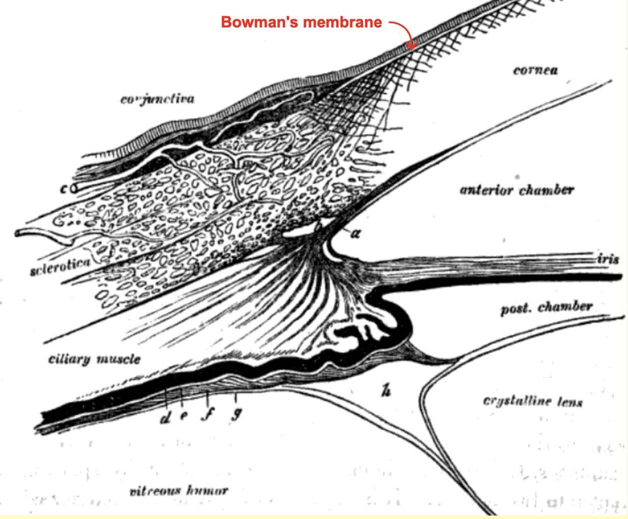
Apart from imaging technology (i.e., drawing rather than photography), Bowman's illustration of the limbus (above) appears practically modern (cf, anterior eyeball, thumbnail image at right). This image is from p. 52 of Bowman's 1847 "Lectures on the parts concerned in the operations on the eye..." [1].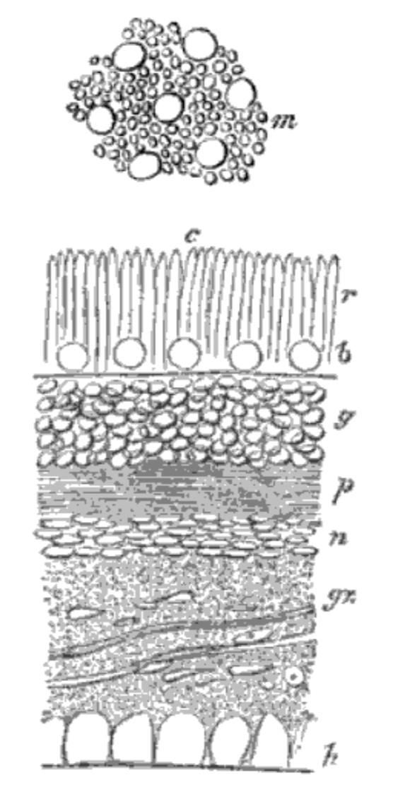
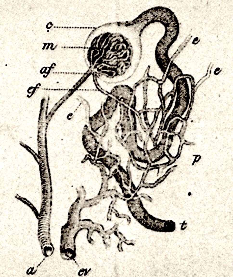
In contrast to the modernity of Bowman's view of the limbus, Bowman's caption for his sketch of retina (at right, from p. 85 in Bowman's "Lectures..." [1]) reveals his limited histological understanding of nervous tissue. The rod and cone outer segments are nicely drawn (cones here are called "bulbs"). But Bowman labels the outer and inner nuclear layers as layers of "g, globular agglomerated granules" and "n, nummular agglomerated granules," since he had not yet appreciated the unifying concept of cell nuclei in Cell Theory (ca. 1839). He describes the outer plexiform layer simply as "obscurely fibrous," which in fact is how it still appears in routine histological preparations (cf., retina on this website).
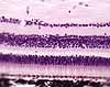
Bowman's upper sketch, labelled m in this illustration, represents a tangential section of rod and cone outer segments in the photoreceptor layer. Bowman's caption identifies this layer as Jacob's membrane, reflecting Bowman's respect for prior work. Although no longer in use today, this term commemorates Irish ophthalmologist Arthur Jacobs, considered to be the first to describe the retina microscopically, in 1819.
Reference cited above
- "Lectures on the parts concerned in the operations on the eye, and on the structure of the retina, delivered at the Royal London Ophthalmic Hospital, Moorfields, June 1847, to which are added, a paper on the vitreous humor; and also a few cases of ophthalmic disease," by William Bowman, F.R.S., Fellow of the Royal College of Surgeons of England; Professor of Physiology and General and Morbid Anatomyin King's Coll. London; Assistant-Surgeon to the King's College Hospital, and to the Royal London Ophthalmic Hospital, Moorfields. Printed for Longman, Brown, Green, and Longmans, 1849. [available at Google Books]
Comments and questions: dgking@siu.edu
SIUC / School
of Medicine / Anatomy / David
King
https://histology.siu.edu/bowman-eye.htm
Last updated: 29 July 2023 / dgk