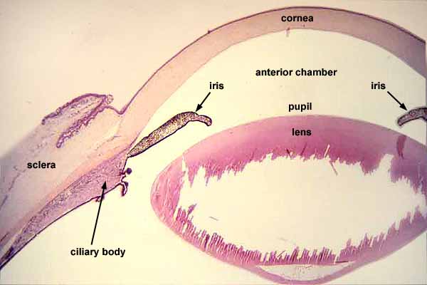
Eye: cornea, iris, lens, and ciliary body

|

| |||||||||||||
For an enlarged view of any of the structures shown here, click on a chosen region within the image, OR click on one of thumbnails below.
NOTE:
Suspensory fibers for the lens are visible only in the enlarged views.
The ragged hole that appears here within the lens is an artifact, the result of difficulty fixing and sectioning this relatively solid tissue.
Comments and questions: dgking@siu.edu
SIUC / School
of Medicine / Anatomy / David
King
https://histology.siu.edu/ssb/EE013b.htm
Last updated: 2 August 2023 / dgk